This site uses cookies to improve your experience. To help us insure we adhere to various privacy regulations, please select your country/region of residence. If you do not select a country, we will assume you are from the United States. Select your Cookie Settings or view our Privacy Policy and Terms of Use.
Cookie Settings
Cookies and similar technologies are used on this website for proper function of the website, for tracking performance analytics and for marketing purposes. We and some of our third-party providers may use cookie data for various purposes. Please review the cookie settings below and choose your preference.
Used for the proper function of the website
Used for monitoring website traffic and interactions
Cookie Settings
Cookies and similar technologies are used on this website for proper function of the website, for tracking performance analytics and for marketing purposes. We and some of our third-party providers may use cookie data for various purposes. Please review the cookie settings below and choose your preference.
Strictly Necessary: Used for the proper function of the website
Performance/Analytics: Used for monitoring website traffic and interactions
Most radiologists have heard of Moore’s Law. A close colleague observed that radiologists are affected by a type of Moore’s Law. The amount of scans a radiologist is expected to shift per day is increasing year on year. Today’s radiologist is expected to shift work at an eye-watering rate. Paul McCoubrie, MBBS.
A significant percentage of imaging studies ordered by office-based healthcare providers are self-interpreted rather than referred to radiologists for reading, according to researchers from the Harvey L. The large differences between radiologists and nonradiologists in interpretation training could lead to differences in diagnostic accuracy."
Out-of-network billing by radiologists – often referred to as “surprise billing” – dropped significantly beginning in 2007 and imaging claims are now almost completely in network, according to a recent study. And by 2021, radiologists practiced almost exclusively in-network, according to the researchers. in 2007 to 1.1%
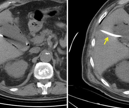
“Based on discussions with our interventional radiology colleagues, dislodged PCNs or severe encrustation may result in much longer and more complex procedures, including the potential need for entirely new access into the collecting system, thereby resulting in higher fluoroscopy use,” Lyon and colleagues wrote.
Neiman Health Policy Institute ( HPI ) study found that radiologists interpreted 72.1% According to a written study summary released by HPI, market share varied by imaging modality; radiologists interpreted 97.3% of radiology/fluoroscopy (XR), 50.9% of ultrasound. of radiology/fluoroscopy (XR), 50.9% of ultrasound.
Here are 6 more 100%-independent radiologist-owned private practices in the US that are recruiting. Onsite procedures include general fluoroscopy, minor ultrasound, paracentesis, and thoracentesis. Onsite procedures include general fluoroscopy, minor ultrasound, paracentesis and thoracentesis. No neuro or MSK.
While conventional chest Xrays and ultrasound can offer clues, additional imaging and tests are often needed for a precise diagnosis, says Michaela Cellina, Head of Imaging Research and a radiologist with ASST Fatebenefratelli Sacco. Diagnosing diaphragm dysfunction is challenging due to its varied symptoms and causes.
The new imaging systems that will be on display include three new digital radiography (DR) suites, two new fluoroscopy systems, a 0.4T Persona C-HR: Fujifilm’s newest mobile fluoroscopy c-arm solution providing 30 frames per second FPS pulsed fluoroscopy images at low dose. The sturdy patient table features a robust 800 lbs.
The Radiology Experience Tour expanded this year to include a portfolio of Philips imaging solutions including Magnetic Resonance, Radiology and Fluoroscopy, Ultrasound, Radiology Operations Command Center (ROCC), PACS, C-Arms and Ambient Experience. Philips also showcased its latest portable ultrasound system, the Compact 5000.
It is crucial to regularly observe various reports like MRI, CT scan, X-Ray, Doppler, Fluoroscopy etc. The description should be vivid enough that any radiologist can understand the intricacies of your findings without having to consult the image or report. Diagnostic Ultrasound- Carol M Rumack Specialized books on neuroradiology i.
Ultrasound Imaging for Soft Tissue Injuries: Real-Time Visualization: Explore the role of ultrasound in imaging soft tissue injuries. Discuss how real-time visualization with ultrasound aids in assessing muscle, tendon, and ligament injuries, providing immediate feedback.
Radiological Milestones: Discuss key milestones in the history of radiology, from the advent of fluoroscopy to the development of advanced imaging techniques. Diverse Imaging Techniques: Explore the wide array of radiological imaging techniques, from X-rays and CT scans to MRI, ultrasound, and nuclear medicine.
Most NPP interpretations were for radiography/fluoroscopy (53.3%) or ultrasound (26.1%). We provide access to a team of board-certified radiologists available around the clock, ensuring timely and accurate interpretations of diagnostic imaging studies. NPP-billed interpretation claims increased from 2.6% in 2016 to 3.3%
These services include X-rays, ultrasounds , MRIs, CT scans, mammography, and fluoroscopy, among others. Staffing and Workforce Management: Recruiting and retaining skilled radiologists, technicians, and support staff is crucial for maintaining quality and efficiency.
For communities in Northern Virginia, low cost, high-quality medical imaging is as close as North Stafford. We are reopening in response to the flattening curve in the Fredericksburg area, and we are doing so in accordance with the CDC, Virginia Department of Health, ACR, and CMS guidelines.
Standardized guidelines can help radiologists navigate the legal landscape of imaging pregnant women with ionizing radiation, according to a July 15 editorial published in the Journal of the American College of Radiology ( ACR ). The same goes for CT imaging outside the abdomen and the pelvic/hip area.
Ultrasound model predicts liver disease progression. Appropriateness and imaging outcomes of ultrasound, CT, and MR in the emergency department: a retrospective analysis from an urban academic center. Radiologist Workforce Attrition from 2019 to 2024: A National Medicare Analysis. SPECT/CT reveals heart’s response to tafamidis.
The patient is pregnant (orange arrows), therefore ionizing radiation with CT scan or fluoroscopy can not be used for imaging guidance. Ultrasound of gallbladder used for guidance of percutaneous needle (red arrow) placement for cholecystostomy. Ultrasound of gallbladder demonstrating drainage catheter in the lumen (blue arrow).
We organize all of the trending information in your field so you don't have to. Join 5,000 users and stay up to date on the latest articles your peers are reading.
You know about us, now we want to get to know you!
Let's personalize your content
Let's get even more personalized
We recognize your account from another site in our network, please click 'Send Email' below to continue with verifying your account and setting a password.


Let's personalize your content