This site uses cookies to improve your experience. To help us insure we adhere to various privacy regulations, please select your country/region of residence. If you do not select a country, we will assume you are from the United States. Select your Cookie Settings or view our Privacy Policy and Terms of Use.
Cookie Settings
Cookies and similar technologies are used on this website for proper function of the website, for tracking performance analytics and for marketing purposes. We and some of our third-party providers may use cookie data for various purposes. Please review the cookie settings below and choose your preference.
Used for the proper function of the website
Used for monitoring website traffic and interactions
Cookie Settings
Cookies and similar technologies are used on this website for proper function of the website, for tracking performance analytics and for marketing purposes. We and some of our third-party providers may use cookie data for various purposes. Please review the cookie settings below and choose your preference.
Strictly Necessary: Used for the proper function of the website
Performance/Analytics: Used for monitoring website traffic and interactions
Over 50% of the world's population (4 billion people) have no access to imaging, a number thought to be much higher in rural areas. Healthcare inequality -- and especially imaging inequality -- appears to be insurmountable on the surface and growing worse with each passing year. This is a solvable problem. It is that simple.
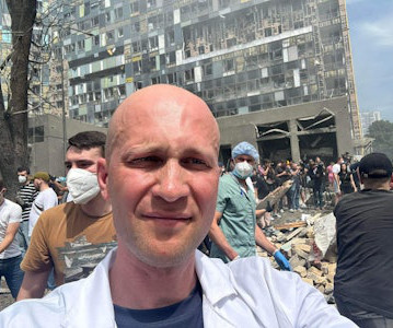
Radiology staff at the Ohmatdyt Children's Hospital in Kyiv are making steady progress with their plan to restore normal service after a devastating missile attack. On July 9, 2024, AuntMinnieEurope.com posted an article about the extensive damage caused to Ukraine's largest pediatric hospital.
is a national platform for imaging AI, Tan explained. Courtesy of Dr Michael Yam, lead for 3D printing centre in Tan Tock Seng Hospital (TTSH). have created anatomical models based on medical images. Imaging-based screening is also an emerging field. AI in healthcare is now a national priority, and AIM.SG
We are pleased to announce GlobalRadiology CME's Kevin Rice, MD is a semifinalist for 2021 ,AuntMinnie.com 's Most Effective Radiology Educator. Rice has authored or co-authored over 200 radiology cases that can be accessed on the GlobalRadiology teaching file.
A: Sagittal CT image demonstrates the right posterior lens dislocation with the lens lying in the dependent portion of the vitreous humor inferiorly (red arrow). B: Sagittal CT image demonstrates the normal location of the left lens in the iris (green arrow). Imaging of orbital trauma. A study of 166 hospitalized cases.
A: Sagittal CT image demonstrates the right posterior lens subluxation with the inferior portion of the lens displaced posteriorly into the vitreous humor (red arrow). B: Sagittal CT image demonstrates the normal location of the left lens in the iris (green arrow). Imaging of orbital trauma. Xray of the Week Figure 1.
Sagittal reformatted CT images. Axial CT imagesImages show horizontal fracture through the right lamina (orange arrows), right pedicle (green arrows) and left pedicle (red arrows). Axial CT image demonstrating the seat belt sign in this patient with stranding in the subcutaneous fat of the abdominal wall (red arrows).
Once the clinical picture and baseline laboratory tests for tumor markers like cancer antigen 15-3 and CEA confirm the diagnosis, local imaging with mammogram and ultrasound will help guide management as standards of care [1]. However, if the lesion is not visualized on imaging, this does not preclude the diagnosis of IBC [1].
During his undergraduate studies, he was involved with multiple volunteer organizations, such as Camp Kemo a summer camp for children with cancer and Palmetto Richland Children’s Hospital. Dr. Rice's passion for state of the art radiology and teaching includes acting as a guest lecturer at UCLA. Follow Dr.
Video going through the axial images on this case. Imaging is important in vasitis as it can prevent unnecessary surgical intervention for other causes of acute groin pain (5-7). Imaging findings prevent unnecessary surgery in vasitis: An under-reported condition mimicking inguinal hernia. Clinical Radiology.
Discussion: The above imaging findings occurred in an 8-year-old child with a trauma after a fall. Radiographically, bowing fractures may show visible bending on radiographic imaging, however, as in this case if the bending occurs within the same plane of the radiograph, there may be no visible deformities on radiographic imaging [2].
More sophisticated imaging, such as computed tomography or magnetic resonance imaging, should be obtained if plain film is unrevealing but there is high suspicion of fracture. Dr. Rice's passion for state of the art radiology and teaching includes acting as a guest lecturer at UCLA. In both 2016 and 2021, Dr.
What are the important findings in each image. A: Coronal CT image demonstrates normal contour of the right globe (green arrow) and a shrunken left globe (orange arrow), which is suggestive of globe rupture. C: Sagittal CT image demonstrates normal contour in the right globe (green arrow). Imaging of orbital trauma.
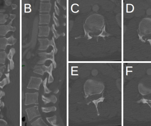
What are the important findings in each image. Coronal CT (A) and sagittal CT (B) images demonstrate fracture at the T11-12 level with significant offset (red arrows) causing the neurologic deficit. Axial CT image (C) demonstrates edema adjacent to the acute fracture (green arrows).
1 ] This diagnostic procedure involves swallowing a pill-sized camera that records thousands of images of the alimentary canal including the small intestine, an area difficult to examine via traditional endoscopy. Rice, MD is the president of GlobalRadiology CME and is a radiologist with Cape Radiology Group.
Imaging and Case Analysis: Radiographic images demonstrate misalignment of the medial side of the second metatarsal with the medial side of the middle cuneiform bone, as seen in this case. Magnetic resonance imaging will help to evaluate ligamentous injury and provides a 94% predictive value for diagnosing Lisfranc injury [5].
X-ray and ultrasound machines were badly damaged in a rocket attack on Ukraine's largest children's hospital on July 8, according to radiologist Stanislav Rebenkov, MD. Rebenkov has been head of radiology at Ohmatdyt Children's Hospital since 2020. Broken glass and damaged equipment are found throughout the hospital.
Like two kids fighting over the same toy, PACS has created a battlefield between radiology and hospital information technology (IT) departments in recent years. Numerous articles have been written about who owns PACS -- radiology or IT -- from both a decision-making and support perspective.
Porencephaly is readily diagnosed via imaging. On CT and MR imaging, porencephalic cysts appear as an intracranial cyst that has a well-defined border and central attenuation the same as CSF (Fig. Obstetric Imaging: Fetal Diagnosis and Care, Elsevier, 2018, pp. Diagnostic Imaging: Obstetrics, Elsevier, 2016, pp.
Diagnosis of CM's is usually an incidental finding on imaging for other indication in asymptomatic patients [4]. Tentative diagnosis is made on imaging upon exclusion of thrombus or vegetation and presence of a mobile mass attached by stalk or a stalk left after mass had embolized systemically [1]. Int J Cardiovasc Imaging.
Urinalysis may show pyuria, leukocytosis, nitrites, hematuria, WBC casts; however, imaging is required to confirm the diagnosis [2,3,4]. Acute gas-producing bacterial renal infection: correlation between imaging findings and clinical outcome. doi: 10.1148/radiology.198.2.8596845 J Microbiol Immunol Infect. 2009;42(5):393-400.
Register now for Imaging in Israel 2023 , then check out all the amazing things to do in Israel! When the sun goes down, enjoy the hospitality of dinner in a Bedouin tent but don't forget to stop to do do a little stargazing before heading to bed. We made a top ten list for some our favorite activities in the holy land.
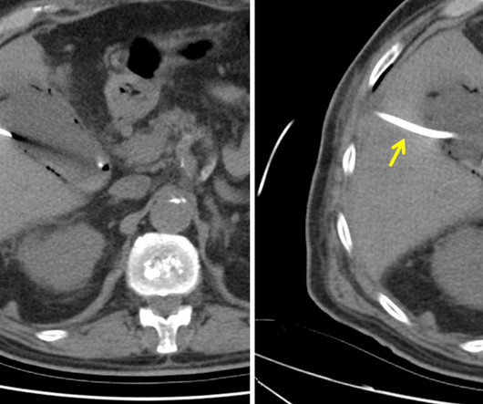
The patient is pregnant (orange arrows), therefore ionizing radiation with CT scan or fluoroscopy can not be used for imaging guidance. The final CT image shows the drainage catheter (yellow arrow) correctly placed in the gallbladder with the tip coiled in the gallbladder fundus (red arrow). MRI of abdomen. Hojberg and Caliskan].
We organize all of the trending information in your field so you don't have to. Join 5,000 users and stay up to date on the latest articles your peers are reading.
You know about us, now we want to get to know you!
Let's personalize your content
Let's get even more personalized
We recognize your account from another site in our network, please click 'Send Email' below to continue with verifying your account and setting a password.


Let's personalize your content