Remove Global Radiology Remove Imaging Remove Radiography Remove Radiology
article thumbnail

Chalk Stick Fracture in Ankylosing Spondylitis

Global Radiology CME

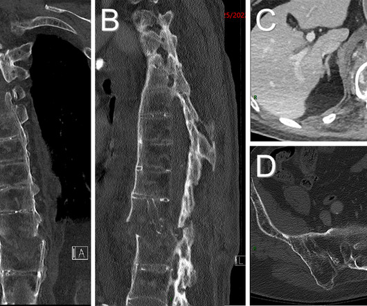
What are the important findings in each image. Coronal CT (A) and sagittal CT (B) images demonstrate fracture at the T11-12 level with significant offset (red arrows) causing the neurologic deficit. Axial CT image (C) demonstrates edema adjacent to the acute fracture (green arrows).

article thumbnail

Lens Subluxation

Global Radiology CME

A: Sagittal CT image demonstrates the right posterior lens subluxation with the inferior portion of the lens displaced posteriorly into the vitreous humor (red arrow). B: Sagittal CT image demonstrates the normal location of the left lens in the iris (green arrow). Imaging of orbital trauma. Xray of the Week Figure 1.

article thumbnail

Lisfranc Fracture Dislocation

Global Radiology CME

Imaging and Case Analysis: Radiographic images demonstrate misalignment of the medial side of the second metatarsal with the medial side of the middle cuneiform bone, as seen in this case. Magnetic resonance imaging will help to evaluate ligamentous injury and provides a 94% predictive value for diagnosing Lisfranc injury [5].