This site uses cookies to improve your experience. To help us insure we adhere to various privacy regulations, please select your country/region of residence. If you do not select a country, we will assume you are from the United States. Select your Cookie Settings or view our Privacy Policy and Terms of Use.
Cookie Settings
Cookies and similar technologies are used on this website for proper function of the website, for tracking performance analytics and for marketing purposes. We and some of our third-party providers may use cookie data for various purposes. Please review the cookie settings below and choose your preference.
Used for the proper function of the website
Used for monitoring website traffic and interactions
Cookie Settings
Cookies and similar technologies are used on this website for proper function of the website, for tracking performance analytics and for marketing purposes. We and some of our third-party providers may use cookie data for various purposes. Please review the cookie settings below and choose your preference.
Strictly Necessary: Used for the proper function of the website
Performance/Analytics: Used for monitoring website traffic and interactions
Over 50% of the world's population (4 billion people) have no access to imaging, a number thought to be much higher in rural areas. Healthcare inequality -- and especially imaging inequality -- appears to be insurmountable on the surface and growing worse with each passing year. This is a solvable problem. It is that simple.
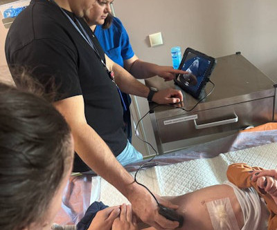
Future healthcare success will rely heavily on giving priority to key technologies like point-of-care ultrasound, 3D printing of anatomical organs, and artificial intelligence, RSNA 2023 attendees are set to find out during Tuesday's keenly anticipated international session on Singapore. is a national platform for imaging AI, Tan explained.
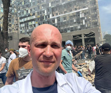
Stanislav Rebenkov, MD, head of radiology, and his team are gradually starting to rebuild the department. Stanislav Rebenkov, MD, has been moved by the support and generosity of the globalradiology community. "On He assisted with the logistics of the delivery process for the Butterfly devices.
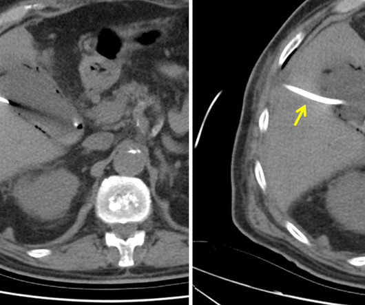
A: Sagittal CT image demonstrates the right posterior lens subluxation with the inferior portion of the lens displaced posteriorly into the vitreous humor (red arrow). B: Sagittal CT image demonstrates the normal location of the left lens in the iris (green arrow). Imaging of orbital trauma. What are the important findings?
Video going through the axial images on this case. Ultrasound of acute vasitis shows a heterogenous and thickened vas deferens. Color Doppler ultrasound of the same section as Figure 4. Imaging is important in vasitis as it can prevent unnecessary surgical intervention for other causes of acute groin pain (5-7).
Ultrasound guided biopsy yielded invasive ductal carcinoma with lymphatic invasion. Once the clinical picture and baseline laboratory tests for tumor markers like cancer antigen 15-3 and CEA confirm the diagnosis, local imaging with mammogram and ultrasound will help guide management as standards of care [1]. Breast Dis.
A: Sagittal CT image demonstrates the right posterior lens dislocation with the lens lying in the dependent portion of the vitreous humor inferiorly (red arrow). B: Sagittal CT image demonstrates the normal location of the left lens in the iris (green arrow). Imaging of orbital trauma. Afr J Emerg Med. 2019;9(2):106-107.
X-ray and ultrasound machines were badly damaged in a rocket attack on Ukraine's largest children's hospital on July 8, according to radiologist Stanislav Rebenkov, MD. Rebenkov said he would welcome any support from the globalradiology community. It will take months to recover, he said. There are broken windows everywhere.
Porencephaly is readily diagnosed via imaging. Antenatal ultrasound may show one or more intracranial cysts that communicate with the ventricular system and/or subarachnoid space. On CT and MR imaging, porencephalic cysts appear as an intracranial cyst that has a well-defined border and central attenuation the same as CSF (Fig.
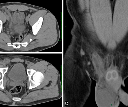
The patient is pregnant (orange arrows), therefore ionizing radiation with CT scan or fluoroscopy can not be used for imaging guidance. Ultrasound of gallbladder used for guidance of percutaneous needle (red arrow) placement for cholecystostomy. Ultrasound of gallbladder demonstrating drainage catheter in the lumen (blue arrow).
We organize all of the trending information in your field so you don't have to. Join 5,000 users and stay up to date on the latest articles your peers are reading.
You know about us, now we want to get to know you!
Let's personalize your content
Let's get even more personalized
We recognize your account from another site in our network, please click 'Send Email' below to continue with verifying your account and setting a password.


Let's personalize your content