This site uses cookies to improve your experience. To help us insure we adhere to various privacy regulations, please select your country/region of residence. If you do not select a country, we will assume you are from the United States. Select your Cookie Settings or view our Privacy Policy and Terms of Use.
Cookie Settings
Cookies and similar technologies are used on this website for proper function of the website, for tracking performance analytics and for marketing purposes. We and some of our third-party providers may use cookie data for various purposes. Please review the cookie settings below and choose your preference.
Used for the proper function of the website
Used for monitoring website traffic and interactions
Cookie Settings
Cookies and similar technologies are used on this website for proper function of the website, for tracking performance analytics and for marketing purposes. We and some of our third-party providers may use cookie data for various purposes. Please review the cookie settings below and choose your preference.
Strictly Necessary: Used for the proper function of the website
Performance/Analytics: Used for monitoring website traffic and interactions
Figure 2 A: AP view radiograph of right forearm. B: Lateral radiograph view of right forearm. An angulated fracture of the distal midshaft radius is also visualized, but there is also bowing of the ulna that is more appreciated on the lateral radiograph view. 8 Year Old Male With Trauma Due To A Fall. Xray of the Week Figure 1.
A) Dorsoplantar radiograph of the foot demonstrating an isolated fracture of the cuboid with possible extension into the tarsometatarsal joint. (B) B) Medial oblique radiograph of the foot demonstrating an isolated fracture of the cuboid. Radiographic evidence can support the diagnosis. Trauma in a 8 year old female.
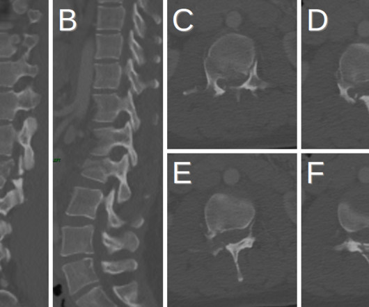
Plain radiograph shows empty vertebral body sign, which results from displacement of the spinous processes [3]. Thoracolumbar Spine Injury at CT: Trauma/Emergency Radiology. Radiographics. 05.0145 Amara Ahmed is a medical student at the ,Florida State University College of Medicine. 2016;36(7):2234-2235. , doi:10.1148/rg.2016160058
A) AP radiograph of Lisfranc Fracture Dislocation demonstrates the circled “fleck sign” or Lisfranc ligament avulsion fracture fragment. (B) C) The lateral radiograph notes with a circle, the dorsal sub dislocation of the metatarsal base. Radiographs should be repeated after two weeks to ensure surgery is unnecessary.
Radiographics. 2015.01.004 Sai Kilaru is a medical student at Central Michigan University College of Medicine and plans to pursue a residency in diagnostic radiology. Sai is also a member of the Gold Humanism Honor Society and is involved with giving back to the community at a local free clinic as a medical assistant.
Frontal abdomen radiograph demonstrates foreign body consistent with capsule endoscopy device (pill cam) in descending colon. A prospective study of the utility of abdominal radiographs after capsule endoscopy for the diagnosis of capsule retention. 61-year-old male with abdominal pain 15 days after capsule endoscopy. doi: 10.3748/wjg.15.2401
Radiographics. doi: 10.1016/0039-6257(82)90069-8 Austin Sanu is a 3rd year medical student at the New York Institute of Technology College of Osteopathic Medicine. He plans on pursuing a residency in Diagnostic Radiology. Dr. Rice's passion for state of the art radiology and teaching includes acting as a guest lecturer at UCLA.
5 Additionally, physicians should avoid maneuvers, including lid retraction and tonometry, or systemic medications that increase intraocular pressure. Radiographics. Through these platforms, he has been instrumental in shaping the future of interventional radiology education and promoting awareness of the field among medical students.
This may include physical therapy, rehabilitation, medication for seizures, shunt, or rarely neurosurgical removal of the cyst. Jay Vora is a medical student at Edward Via College of Osteopathic Medicine (VCOM) – Virginia and plans to pursue a residency in diagnostic radiology. doi: 10.1016/B978-0-443-07416-5.50011-3.
We organize all of the trending information in your field so you don't have to. Join 5,000 users and stay up to date on the latest articles your peers are reading.
You know about us, now we want to get to know you!
Let's personalize your content
Let's get even more personalized
We recognize your account from another site in our network, please click 'Send Email' below to continue with verifying your account and setting a password.


Let's personalize your content