This site uses cookies to improve your experience. To help us insure we adhere to various privacy regulations, please select your country/region of residence. If you do not select a country, we will assume you are from the United States. Select your Cookie Settings or view our Privacy Policy and Terms of Use.
Cookie Settings
Cookies and similar technologies are used on this website for proper function of the website, for tracking performance analytics and for marketing purposes. We and some of our third-party providers may use cookie data for various purposes. Please review the cookie settings below and choose your preference.
Used for the proper function of the website
Used for monitoring website traffic and interactions
Cookie Settings
Cookies and similar technologies are used on this website for proper function of the website, for tracking performance analytics and for marketing purposes. We and some of our third-party providers may use cookie data for various purposes. Please review the cookie settings below and choose your preference.
Strictly Necessary: Used for the proper function of the website
Performance/Analytics: Used for monitoring website traffic and interactions
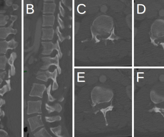
On MRI, there may be a bright T2 signal signifying edema with low signal intensity fracture lines, intervertebral disc injury, and spinal cord edema [1]. Plain radiograph shows empty vertebral body sign, which results from displacement of the spinous processes [3]. Thoracolumbar Spine Injury at CT: Trauma/Emergency Radiology.
On MRI, the cystic components will appear as low signal intensity on T1 weighted images, high signal intensity on T2 weighted images, low signal intensity on FLAIR images, and will have no restricted diffusion on DWI [10-12]. He discovered his passion for radiology during the first radiology lecture at VCOM.
We organize all of the trending information in your field so you don't have to. Join 5,000 users and stay up to date on the latest articles your peers are reading.
You know about us, now we want to get to know you!
Let's personalize your content
Let's get even more personalized
We recognize your account from another site in our network, please click 'Send Email' below to continue with verifying your account and setting a password.


Let's personalize your content