Remove Global Radiology Remove Presentation Remove Radiographer
article thumbnail

Lisfranc Fracture Dislocation

Global Radiology CME

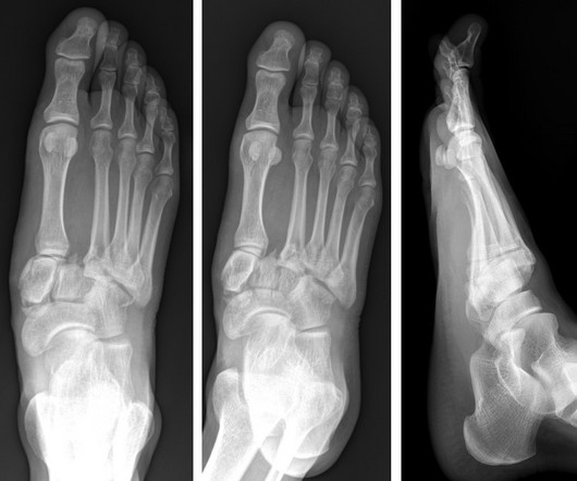
A) AP radiograph of Lisfranc Fracture Dislocation demonstrates the circled “fleck sign” or Lisfranc ligament avulsion fracture fragment. (B) C) The lateral radiograph notes with a circle, the dorsal sub dislocation of the metatarsal base. Trauma due to falling off a roof. Xray of the Week Figure 1. Trauma due to falling off a roof.

article thumbnail

Plastic Bowing Fracture Ulna

Global Radiology CME

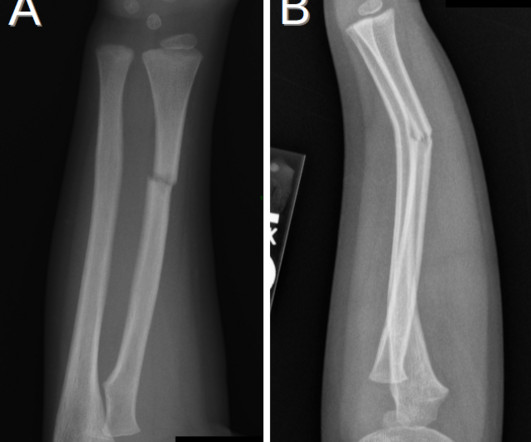
Figure 2 A: AP view radiograph of right forearm. B: Lateral radiograph view of right forearm. An angulated fracture of the distal midshaft radius is also visualized, but there is also bowing of the ulna that is more appreciated on the lateral radiograph view. 8 Year Old Male With Trauma Due To A Fall. Xray of the Week Figure 1.

Insiders

Sign Up for our Newsletter

This site is protected by reCAPTCHA and the Google Privacy Policy and Terms of Service apply.

Trending Sources

article thumbnail

Chance fracture

Global Radiology CME

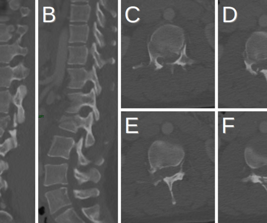
Fracture of the spinous process (blue arrows) is also present. Chance fractures are difficult to identify as they do not present with neurological deficits but can present with intra-abdominal injuries [1]. Plain radiograph shows empty vertebral body sign, which results from displacement of the spinous processes [3].

article thumbnail

Lens Subluxation

Global Radiology CME

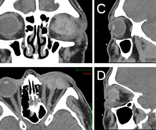
Patients with ectopia lentis typically present with visual acuity problems but can also present with eye pain if secondary to trauma [2]. Anterior axial lens subluxation, progressive myopia, and angle closure glaucoma: recognition and treatment of atypical presentation of ectopia lentis. Radiographics. 2006;10(4):345-350.

article thumbnail

Emphysematous Pyelonephritis

Global Radiology CME

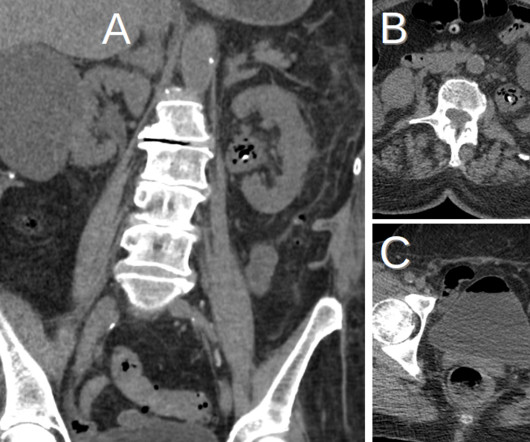
Like pyelonephritis, patients with EPN often present with fever, abdominal or flank pain, and costovertebral angle tenderness. KUB indicates kidneys, ureter, and bladder (plain abdominal radiograph); CT, computed tomography; and PCD, percutaneous catheter drainage. Pyelonephritis: Radiologic-Pathologic Review. RadioGraphics.

article thumbnail

Ruptured Globe

Global Radiology CME

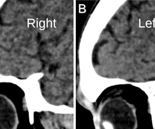
Thin-section axial CT scans followed by multiplanar reformations can visualize ruptured globe contour, which can present as a “mushroom” (Figs. The patient’s presenting visual acuity is a major determinant of their post-surgical visual acuity. Radiographics. 1-2) or “flat tire” shape. Clin Ophthalmol. 2013;7:461-465.

article thumbnail

Porencephaly

Global Radiology CME

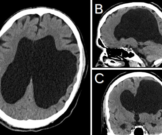
The earlier the insult occurred in development, the more likely a glial reaction will be present. He discovered his passion for radiology during the first radiology lecture at VCOM. Seeing the radiographic images made medical education come to life for him. Follow Jay Vora on Twitter @JS_Vora ,Kevin M.