This site uses cookies to improve your experience. To help us insure we adhere to various privacy regulations, please select your country/region of residence. If you do not select a country, we will assume you are from the United States. Select your Cookie Settings or view our Privacy Policy and Terms of Use.
Cookie Settings
Cookies and similar technologies are used on this website for proper function of the website, for tracking performance analytics and for marketing purposes. We and some of our third-party providers may use cookie data for various purposes. Please review the cookie settings below and choose your preference.
Used for the proper function of the website
Used for monitoring website traffic and interactions
Cookie Settings
Cookies and similar technologies are used on this website for proper function of the website, for tracking performance analytics and for marketing purposes. We and some of our third-party providers may use cookie data for various purposes. Please review the cookie settings below and choose your preference.
Strictly Necessary: Used for the proper function of the website
Performance/Analytics: Used for monitoring website traffic and interactions
Figure 2 A: AP view radiograph of right forearm. B: Lateral radiograph view of right forearm. An angulated fracture of the distal midshaft radius is also visualized, but there is also bowing of the ulna that is more appreciated on the lateral radiograph view. 8 Year Old Male With Trauma Due To A Fall. Xray of the Week Figure 1.
A) Dorsoplantar radiograph of the foot demonstrating an isolated fracture of the cuboid with possible extension into the tarsometatarsal joint. (B) B) Medial oblique radiograph of the foot demonstrating an isolated fracture of the cuboid. Radiographic evidence can support the diagnosis. Trauma in a 8 year old female.
A) AP radiograph of Lisfranc Fracture Dislocation demonstrates the circled “fleck sign” or Lisfranc ligament avulsion fracture fragment. (B) C) The lateral radiograph notes with a circle, the dorsal sub dislocation of the metatarsal base. Radiographs should be repeated after two weeks to ensure surgery is unnecessary.
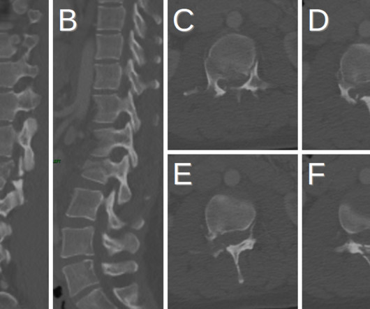
Plain radiograph shows empty vertebral body sign, which results from displacement of the spinous processes [3]. Thoracolumbar Spine Injury at CT: Trauma/Emergency Radiology. Radiographics. Dr. Rice's passion for state of the art radiology and teaching includes acting as a guest lecturer at UCLA. 2016;36(7):2234-2235. ,
A) AP radiograph demonstrates the talus (yellow arrow) with anatomical alignment to the distal tibia. B) Lateral radiograph demonstrates the talus (yellow arrow) with anatomical alignment The patient jumped to catch a baseball and landed while rotating to the right. Lateral Subtalar Joint Dislocation. (A)
Radiographics. Rice, MD is the president of GlobalRadiology CME Dr. Rice is a radiologist with ,Renaissance Imaging Medical Associates and is currently the Vice Chief of Staff at ,Valley Presbyterian Hospital in Los Angeles, California. 2006;10(4):345-350. doi: 10.1016/j.jaapos.2006.01.218 2006.01.218 Kubal WS. doi: 10.1148/rg.286085523
Frontal abdomen radiograph demonstrates foreign body consistent with capsule endoscopy device (pill cam) in descending colon. A prospective study of the utility of abdominal radiographs after capsule endoscopy for the diagnosis of capsule retention. 61-year-old male with abdominal pain 15 days after capsule endoscopy. doi: 10.3748/wjg.15.2401
Radiographics. Rice, MD is the president of GlobalRadiology CME Dr. Rice is a radiologist with ,Renaissance Imaging Medical Associates and is currently the Vice Chief of Staff at ,Valley Presbyterian Hospital in Los Angeles, California. Afr J Emerg Med. 2019;9(2):106-107. doi: 10.1016/j.afjem.2019.01.001 Am J Optom Physiol Opt.
Radiographics. Rice, MD is the president of GlobalRadiology CME and is a radiologist with Cape Radiology Group. Dr. Rice's passion for state of the art radiology and teaching includes acting as a guest lecturer at UCLA. Clinical features of single and repeated globe rupture after penetrating keratoplasty.
KUB indicates kidneys, ureter, and bladder (plain abdominal radiograph); CT, computed tomography; and PCD, percutaneous catheter drainage. Pyelonephritis: Radiologic-Pathologic Review. RadioGraphics. Dr. Rice's passion for state of the art radiology and teaching includes acting as a guest lecturer at UCLA.
He discovered his passion for radiology during the first radiology lecture at VCOM. Seeing the radiographic images made medical education come to life for him. While shadowing and on rotations, Jay saw how integral the field of radiology is to every other specialty in medicine and its key role in patient care.
We organize all of the trending information in your field so you don't have to. Join 5,000 users and stay up to date on the latest articles your peers are reading.
You know about us, now we want to get to know you!
Let's personalize your content
Let's get even more personalized
We recognize your account from another site in our network, please click 'Send Email' below to continue with verifying your account and setting a password.


Let's personalize your content