This site uses cookies to improve your experience. To help us insure we adhere to various privacy regulations, please select your country/region of residence. If you do not select a country, we will assume you are from the United States. Select your Cookie Settings or view our Privacy Policy and Terms of Use.
Cookie Settings
Cookies and similar technologies are used on this website for proper function of the website, for tracking performance analytics and for marketing purposes. We and some of our third-party providers may use cookie data for various purposes. Please review the cookie settings below and choose your preference.
Used for the proper function of the website
Used for monitoring website traffic and interactions
Cookie Settings
Cookies and similar technologies are used on this website for proper function of the website, for tracking performance analytics and for marketing purposes. We and some of our third-party providers may use cookie data for various purposes. Please review the cookie settings below and choose your preference.
Strictly Necessary: Used for the proper function of the website
Performance/Analytics: Used for monitoring website traffic and interactions
While services for breast and lung cancer screening were temporarily halted, imagers in x-ray, lung ultrasound, and PET/CT were busy examining patients who presented with COVID-19. Her team employed a tracking mechanism for patients who were due for their mammograms once screening operations resumed.
Maybe you recently decided to try the best 3D mammogram experience in El Paso, have recently moved, changed doctors, or acquired new insurance, and are now going to our imaging center for your annual mammogram. In this case, the radiologist may recommend a diagnostic mammogram. So, why are these prior images so important?
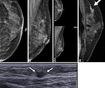
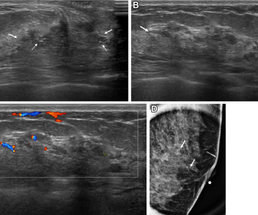
A team led by Su Min Ha, MD, PhD, from Seoul National University Hospital in South Korea reported that AI by itself achieved a higher performance than radiologists with no AI help when it came to detecting contralateral breast cancer in women treated with unilateral mastectomy. years after right mastectomy. (A)
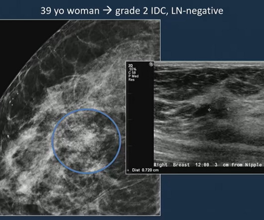
Images of a 39-year-old woman shows a grade 2 invasive ductal carcinoma on mammography, confirmed by supplementary ultrasound. Mammography detected an asymmetry with calcifications while ultrasound examination revealed a hyperechoic mass. Additionally, all exams performed from 2013 to 2019 included digital breast tomosynthesis (DBT).
Previous studies have demonstrated that dense breast tissue masks breast cancers on mammography, and that supplemental imaging such as ultrasound and MRI confirms suspicious findings within dense tissue. Radiologists have studied and continue to research how breast density plays into breast cancer risk. The study can be found here.
The reduction for the bilateral mammogram 77066 was 1.36%, reflecting an increase in RVU valuation that somewhat offsets the conversion factor cut. Effect on professional component reimbursement The single-view chest x-ray 71045 professional fee was cut 5.55%. Overall, the professional component reimbursement is estimated to decrease 2.7%
A team led by Julie Hamzah, MBBS, from Singapore General Hospital, found that symptomatic first breast cancers, dense breasts, and the presence of trabecular thickening on mammography are tied to mammogram detection failure of ipsilateral second breast cancers.
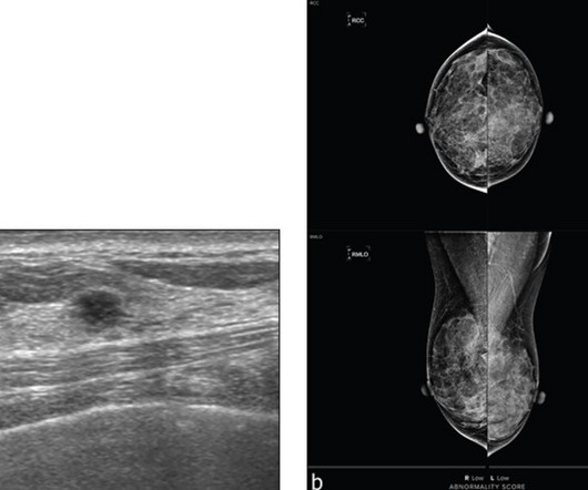
milla1cf Fri, 07/28/2023 - 23:31 July 28, 2023 — Findings from an accepted manuscript published in the American Journal of Roentgenology (AJR) suggest that for patients with dense breasts undergoing screening in the incidence setting, a commercial AI tool did not provide additional benefit to mammography with supplementary ultrasound (US).
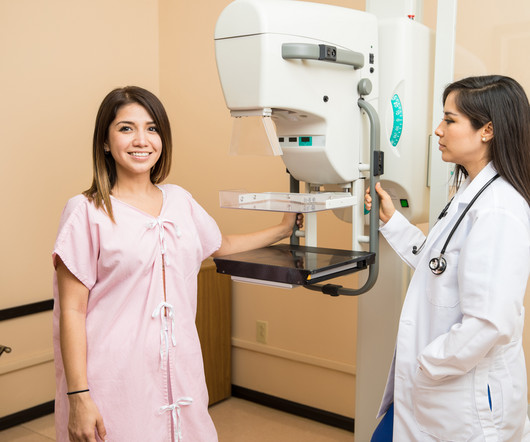
Capitol Imaging Services has served tens of thousands of women with our women’s health examinations in screening mammography, diagnostic mammography, ultrasound, bone density studies, breast MRI and breast biopsy. For screening mammograms, we ask for about 20 minutes of a woman’s time.
Maybe you have recently moved, changed doctors, or have new insurance and now, are going to a different imaging center for your annual mammogram. Either way, the new imaging center will very often request that you provide the images from your previous mammograms. In this case, the radiologist may recommend a diagnostic mammogram.
milla1cf Mon, 07/31/2023 - 21:28 July 31, 2023 — Findings from an accepted manuscript published in the American Journal of Roentgenology (AJR) suggest that for patients with dense breasts undergoing screening in the incidence setting, a commercial AI tool did not provide additional benefit to mammography with supplementary ultrasound ( US ).
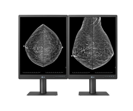
Suitable for reviewing various types of medical images, including breast MRIs, CT scans and ultrasound, the diagnostic monitor features an internal front sensor that removes the need for an external calibration device. LG’s expanded lineup of diagnostic monitors covers the major display needs of large hospitals.
As more hospitals and imaging facilities adopt iCAD’s Breast AI Suite, customers increasingly report the Company’s technologies offer a range of benefits beyond cancer detection,” said Ms. Our ROI tool enables prospective customers to envision how an investment in our ProFound AI solution can pay off for clinicians, facilities, and patients.”
It might be an MRI, an ultrasound, a mammogram, or another imaging test. When a primary care doctor orders specialized testing, say for a patient who complains of breast pain, they may not know the best imaging test to choose.
With an X-ray, Ultrasound, Mammogram and CT scan at our disposal, we needed to have a radiologist who could read all these modalities, and give us results in the shortest time possible to enable us to give the best medical care possible” she says. MHRG devised a 24/7 remote radiology solution for the Clinic.
At Manipal Hospitals Radiology Group, we believe in reaching out to the remotest corners and providing quality radiology reports which make a difference. Mutema’s vision, she entrusted the responsibility of making this care accessible to her patients to Manipal Hospitals Radiology Group (MHRG).
Medical Imaging of Fredericksburg’s services include X-Ray, CT, PET/CT, MRI, 3D mammograms, Ultrasound and a variety of other health scans. Patients have given an over 95% satisfaction score – a reflection of the level of commitment the physicians and staff have to the community they serve.
A) Mammogram MLO view. Mammogram CC view. A mammogram demonstrated focal asymmetries involving most of the anterior and mid right breast with diffuse skin thickening, trabecular coarsening, increased overall density, and enlarged right axillary lymph nodes. What is the diagnosis? Xray of the Week Figure 1.
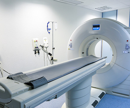
There are several types of imaging tests that physicians use to detect cancer in patients: X-Ray, Computed Tomography (CT), Magnetic Resonance Imaging (MRI), Ultrasound (US), Nuclear Medicine, and Positron Emission Tomography (PET). Are Imaging Tests for Cancer only Available at Hospitals?
Our radiologists with breast imaging expertise can re-evaluate mammograms, breast MRIs, and ultrasounds. Ultrasound Obstetric and Gynecologic Ultrasounds : Subtle findings such as ovarian cysts or fetal abnormalities often require expert review to confirm a diagnosis.
All of the annual scheduled services such as mammograms can now be scheduled, as well as imaging prescribed by physicians for the care of their patients. Patients may also schedule mammograms directly at our facilities in these same locations. What this means for our community and region is important.
Hence, mammograms carried out anywhere can now be viewed by expert breast radiologists in any part of the world thanks to teleradiology. If the breast is dense on the mammogram, an ultrasound must also be carried out. With the advancement in technology and the world going digital, mammography machines are now digital.
a screening mammogram). With locations from Texas to Florida, getting the highest-quality MRI, CT, ultrasound or other type of exam is convenient and yet may cost much less. Ask about your options for free preventative care. Check with your HR department or your doctor about what care is available to you.
What’s more, the USPSTF concluded that there was insufficient evidence to recommend supplemental screening with MRI or ultrasound in women, regardless of breast density. Food and Drug Administration (FDA) requirement that all women having mammograms receive notice that their breasts are dense or not dense.
Within weeks of his announcement hospitals world-wide had taken the initiative to open up X-ray rooms, which gave rise to the first radiology departments. (3) Portrait of Sir Godfrey Hounsfield (1919-2004) The first clinical CT scan: Atkinson Morley's Hospital, October 1971 Credit: impactscan.org.
Some nonmass breast lesion characteristics on ultrasound exams can be considered suspicious for malignancy at screening ultrasound, according to research published November 5 in Radiology. C) Longitudinal color Doppler ultrasound image shows high vascularity. (D) vs. 24% to 55%) than having a positive mammogram.
We organize all of the trending information in your field so you don't have to. Join 5,000 users and stay up to date on the latest articles your peers are reading.
You know about us, now we want to get to know you!
Let's personalize your content
Let's get even more personalized
We recognize your account from another site in our network, please click 'Send Email' below to continue with verifying your account and setting a password.


Let's personalize your content