This site uses cookies to improve your experience. To help us insure we adhere to various privacy regulations, please select your country/region of residence. If you do not select a country, we will assume you are from the United States. Select your Cookie Settings or view our Privacy Policy and Terms of Use.
Cookie Settings
Cookies and similar technologies are used on this website for proper function of the website, for tracking performance analytics and for marketing purposes. We and some of our third-party providers may use cookie data for various purposes. Please review the cookie settings below and choose your preference.
Used for the proper function of the website
Used for monitoring website traffic and interactions
Cookie Settings
Cookies and similar technologies are used on this website for proper function of the website, for tracking performance analytics and for marketing purposes. We and some of our third-party providers may use cookie data for various purposes. Please review the cookie settings below and choose your preference.
Strictly Necessary: Used for the proper function of the website
Performance/Analytics: Used for monitoring website traffic and interactions
When I was in short medical trousers, I grew up surrounded by mighty plain film gods. Some of my radiological heroes would report a staggering 30,000 to 40,000 radiographs a year. Some even [startled gasp] gave up reporting plain radiographs. Not doubling every two years but expectations are steadily growing.
He earned his medical degree at the University of Alabama School of Medicine and completed a residency in diagnostic radiology at the University of Florida. I'm a radiographer,' " Stewart recalled. My aha moment started in the fall of 2015 when I realized that informatics played such a huge role in medical imaging," Stewart said. "I
The finding by researchers in Copenhagen, Denmark, suggests that AI could eventually help streamline high-volume radiology workflows by handling some of the more “tedious parts of the work,” lead author Louis Plesner, MD, of Herlev and Gentofte Hospital in Denmark told AuntMinnie.com.
SINGAPORE - AI is already changing the practice of medical imaging, and there's more technological growth to come, according to a plenary talk delivered May 4 at the International Society of Magnetic Resonance in Medicine (ISMRM) annual meeting. Food and Drug Administration (FDA) were for radiology indications.
He earned his medical degree at the University of Alabama School of Medicine and completed a residency in diagnostic radiology at the University of Florida. Pickhardt serves as chief of gastrointestinal imaging at the University of Wisconsin in Madison and is medical director of oncological imaging at the UW Carbone Cancer Center.
Gleamer) increased sensitivity for detecting all abnormalities on x-rays for all readers (thoracic radiologists, general radiologists, and radiology residents), according to a group of Gleamer consultants and clinicians at Cochin Hospital in Paris. In a retrospective study, a commercially available algorithm (ChestView, v.
ChatGPT can answer patient questions about radiation protection for medical imaging exams comparably to websites of radiology institutions, according to research published June 25 in Radiology. and Europe to evaluate both sets of answers, blinded to the source.
The Karolinska hospital group has recently established an MR safety team involving staff at Solna and Huddinge. The Karolinska hospital group has recently established an MR safety team involving staff at Solna and Huddinge. The tanks are normally color-coded for nonmagnetic material, but this tank had been labeled incorrectly.
Presenter Yoichi Sato, MD, of Tokyo Shinjuku Medical Center in Japan, and colleagues first developed the model using patient dual-energy x-ray absorptiometry (DEXA) T-scores (bone mineral density) and chest x-rays as input. The model achieved 79% accuracy, 96.6% sensitivity, and 34.1% specificity in predicting T-scores ≤ -1.0 accuracy, 77.1%
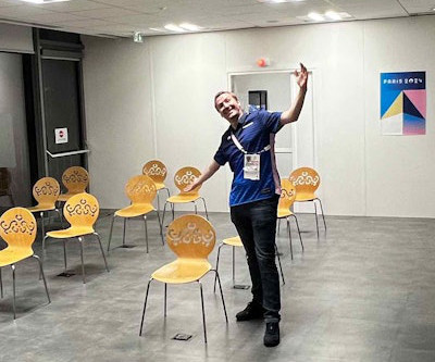
The first scans have been performed in the Olympic imaging polyclinic ahead of Friday's opening ceremony, and the 68-strong squad of radiologists and radiographers are primed and ready for action, according to musculoskeletal (MSK) expert Jérôme Renoux, MD. Bring it on!
“This is a potentially practice-changing trial,” said Oliver Sartor, MD, a medical oncologist and director of radiopharmaceutical trials at the Mayo Clinic in Rochester, MN. He presented the abstract on behalf of first author Ken Herrmann, MD, chair of nuclear medicine at the University Hospital Essen in Essen, Germany.
A team led by Junqi Han, MD, from the Affiliated Hospital of Qingdao University in China found that its model combining data from mammography images, ultrasound images, and other characteristics performed well in predicting disease-free survival of breast cancer.
Significantly, however, AI assistance allowed participants to cut reading times by nearly half, noted lead author Lili Guo, MD, of Nanjing Medical University, and colleagues. The study was published February 8 in the Journal of Imaging Informatics in Medicine.
Reading Time: 10 minutes read By Henry Williams, Carestream Area Vice President, Sales Western Nowadays, with hospital budgetary restrictions at the forefront of the purchasing decision making process, it seems like the X-Ray market, like everything else, is not immune to the current state of the economy. Who is Making the Purchases?
In an open forum, Yi Xiang Tay, of Singapore University Hospital's radiography and diagnostic imaging department, shared his team's research. Also, referral guidelines contributed to more consistent medical care which translated into patient satisfaction, he said.
Perry Pickhardt, MD, UW-Madison While Pickhardt’s efforts revolve around what is commonly called “opportunistic screening,” he likens the goals to value-based medical imaging and repurposing scans for useful incidental data for cardiometabolic screening purposes. Cooky’s tough love: It takes time to become an excellent radiologist.
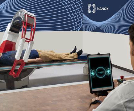
an innovative medical imaging technology company, today announced that it has received a 510(k) clearance from the U.S. Medical imaging systems are an important early detection tool that are key to initiating early treatment, improving health outcomes, and ultimately saving lives.
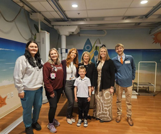
Reading Time: 5 minutes read The following is intended for promotional purposes only and is not medical advice. Earlier this spring, Carestream’s Digital Marketing Manager, John Crowther, had the opportunity to travel to Shriner’s Children’s Hospital in St. Working in healthcare comes with a sense of pride and purpose.
The American Society of Radiologic Technologists (ASRT) has partnered with medical imaging professional and actor, Michael Benzaia, for its new "Be Seen" campaign to raise public awareness about the role of medical imaging and radiation therapy professionals in patient diagnosis, intervention, and treatment.
If you work in radiology in a community healthcare facility, you may have low demand for pediatric medical imaging. I am passionate about improving the performance and interpretation of pediatric medical imaging. The balance of dose and image quality is even more important in pediatric medical imaging.
11, 2025 Harrison.ai, a developer of AI-powered medical diagnostic support and workflow solutions, has announced the accelerated expansion of its operations into the United States a move supported by US$112 million of Series C funding. Averaged across all findings on chest radiographs.) [2]AIDE tim.hodson Wed, 02/12/2025 - 09:30 Feb.
A team of 32 radiologists and 36 radiographers are limbering up to work at the summer Olympics, which begins July 25. Renoux and Crema both attended the Tokyo Olympics, working as part of the medical staff of the French Olympic team. If they need more acute care, they will be referred to the local hospital,” he explained.
The white paper evaluates the capabilities of Canon Intelligent NR and its deep learning neural network (DLNN) software in relation to standard and decreased dose pediatric digital radiographs at Dayton Children’s Hospital, Dayton, Ohio.
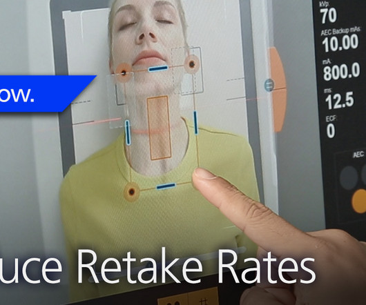
Repeating imaging exams increases the workload of your radiographers who are already stretched too thin; increases the exposure of the affected patients; and contributes to patients’ reduced confidence and satisfaction with your imaging department. The Audio Assist makes it easier for radiographers to hear the patients.
Additionally, all table movements can be controlled by the radiographer from the touch screen, enabling easy setup of either fluoroscopy or radiography settings. A camera integrated into the collimators allows the radiographer to position the patient directly from the console without using fluoroscopy.
This product is a collaboration with UMG/Del Medical. Reveal’s ability to simultaneously acquire conventional and dual-energy images with a single exposure at the bedside improves hospital and patient outcomes and protects revenue by reducing outflows. Our radiographic spectral images separate materials such as water (i.e.,
milla1cf Thu, 11/23/2023 - 06:00 November 23, 2023 — Fujifilm Healthcare Americas Corporation, a leading provider of diagnostic and enterprise imaging solutions, is unveiling several new medical systems at the 2023 Radiological Society of North America ( RSNA ) annual meeting, booth #1929, held November 26 – 30 at McCormick Place in Chicago.
milla1cf Fri, 02/23/2024 - 10:22 February 23, 2024 — The American Society of Radiologic Technologists (ASRT) launched its "Be Seen" campaign today to raise public awareness about the crucial role medical imaging and radiation therapy professionals play in patient diagnosis, intervention and treatment.
The enduring shortage is affecting staffing for radiographers and radiologists; and all imaging modalities. Travel positions offer better pay while the increasing number of urgent care and free-standing emergency centers offer an environment that is more attractive and less stressful to many candidates than a hospital.
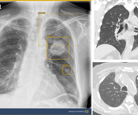
Chest radiography is the most common medical imaging tool used in routine clinical practices to identify different disease findings. Augmento X-Ray represents a significant breakthrough in AI-powered medical imaging," said Ajit Patil, Co-Founder of DeepTek. billion annual X-rays performed, 1.5 With its operations in Pune; DeepTek.ai
Key Points: Currently plain radiographs are the standard method in diagnosing syndesmotic ankle injuries even though the distal tibiofibular joint cannot be assessed due to superposition of the osseous structures in the foot. Dr. Peiffer et.
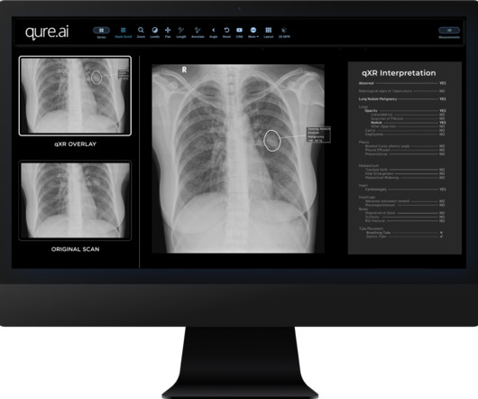
christine.book Tue, 09/12/2023 - 11:56 September 12, 2023 — Medical imaging Artificial Intelligence (AI) company Qure.ai Digumarthy, MD , a senior co-author of the study and thoracic radiologist at Massachusetts General Hospital (Boston, MA), reported up to 96% sensitivity and 100% specificity for the qXR algorithm.
milla1cf Thu, 08/17/2023 - 11:51 August 17, 2023 — University of Missouri School of Medicine neurologist Adnan Qureshi, MD recently led a study that discovered last year’s iodinated media contrast dye shortage affected the assessment of stroke patients at hospitals across the country.
The product, which is a collaboration with UMG/Del Medical, will be showcased at the upcoming AHRA Annual Meeting. This partnership with Del Medical comes at an excellent time,” said Amol Karnick , President and CEO of KA Imaging. The Reveal 35C detector mimics the workflow, dose and techniques of state-of-the-art mobile DR X-ray.
Researchers found that first-ray hypermobility primarily arises from TMT1 instability, though there is still a limited understanding of TMT1 morphology and anatomy due to the challenges and limitations of radiographic imaging.
Does it support new medical imaging software that can help improve clinical outcomes? Physicians have better diagnostic confidence when they can view radiographs in the manner most suitable to their preferences. Your radiographers need to work as efficiently as possible in order to keep pace.
Introduction: In the digital era of healthcare, Picture Archiving and Communication Systems (PACS) have emerged as orchestras orchestrating the seamless management and communication of medical images. Efficient Image Storage and Retrieval: Explore how PACS facilitates efficient storage and retrieval of medical images.
Specializing in abdominal radiology, Dr. Lee is chief of women’s imaging and officer of mentored research in the Department of Radiology at Massachusetts General Hospital ( MGH ) and associate professor of radiology at Harvard Medical School in Boston. “I She has been the editor of RSNA’s Radiology In Training since 2019.
Sites: Investigators recruited patients at 31 French emergency departments at university and nonuniversity hospitals Duration : June 1, 2009 to March 31, 2015. Funding: The Cardinal Health Laboratory provided Turkel thoracentesis kits to two hospitals Trial Registry: Clinicaltrials.gov: NCT01008228.
Kim Mason Kim Mason, an Audit and Research Radiographer for Mid Yorkshire Teaching Hospitals Trust, talks about their role as well as the value of radiographer engagement in research activities and how to get involved. So, what is an Audit and Research Radiographer? All beginning with that first piece of research.
Figure 2 A: AP view radiograph of right forearm. B: Lateral radiograph view of right forearm. An angulated fracture of the distal midshaft radius is also visualized, but there is also bowing of the ulna that is more appreciated on the lateral radiograph view. 8 Year Old Male With Trauma Due To A Fall. Xray of the Week Figure 1.
Secondary Safety Outcomes: The frequency of side effects related to treatment, anxiety, and depression was evaluated with the Hospital Anxiety and Depression Scale at 1 month and 3 months All-cause mortality RESULTS 50 patients were enrolled in the trial Trial enrollment was discontinued due to the COVID-19 pandemic and the cessation of funding.
Knee osteoarthritis (OA) clinical trials results show that weight bearing CT (WBCT) imaging may offer new insights into OA pathology beyond what plain radiographs and MRI can provide. Tom Turmezei, MPhil, MA, BMBCh, PhD, FRCR, a consultant radiologist at Norfolk and Norwich University Hospitals NHS Foundation Trust in Norfolk, UK.
A) Dorsoplantar radiograph of the foot demonstrating an isolated fracture of the cuboid with possible extension into the tarsometatarsal joint. (B) B) Medial oblique radiograph of the foot demonstrating an isolated fracture of the cuboid. Radiographic evidence can support the diagnosis. Trauma in a 8 year old female.
We organize all of the trending information in your field so you don't have to. Join 5,000 users and stay up to date on the latest articles your peers are reading.
You know about us, now we want to get to know you!
Let's personalize your content
Let's get even more personalized
We recognize your account from another site in our network, please click 'Send Email' below to continue with verifying your account and setting a password.


Let's personalize your content