This site uses cookies to improve your experience. To help us insure we adhere to various privacy regulations, please select your country/region of residence. If you do not select a country, we will assume you are from the United States. Select your Cookie Settings or view our Privacy Policy and Terms of Use.
Cookie Settings
Cookies and similar technologies are used on this website for proper function of the website, for tracking performance analytics and for marketing purposes. We and some of our third-party providers may use cookie data for various purposes. Please review the cookie settings below and choose your preference.
Used for the proper function of the website
Used for monitoring website traffic and interactions
Cookie Settings
Cookies and similar technologies are used on this website for proper function of the website, for tracking performance analytics and for marketing purposes. We and some of our third-party providers may use cookie data for various purposes. Please review the cookie settings below and choose your preference.
Strictly Necessary: Used for the proper function of the website
Performance/Analytics: Used for monitoring website traffic and interactions
TORONTO – The radiopharmaceutical Pluvicto improves progression-free survival in patients with metastatic prostate cancer who haven’t undergone taxane therapy, according to research presented at the SNMMI meeting. Oliver Sartor, MD The study, which was awarded SNMMI’s Abstract of the Year award, shared data from the phase III PSMAfore trial.
Charlene Liew, MD, director of cardiothoracic imaging at Changi General Hospital in Singapore, outlined the current state of AI in radiology and where it is going in a presentation called "AI in Radiology: The Past Informs the Future."
M4-SSMK03-4 | Room E450A A deep-learning AI model will be presented in this session that can predict bone mineral density T-scores from chest x-rays. They then trained the algorithm on a data set of 47,150 x-rays (23,151 patients) and validated it on an external data set of 2,914 radiographs (1,515 patients). 11:40 a.m. | accuracy, 77.1%
Presenting the research on November 28 at RSNA 2023, Jan Rudolph, MD, from the department of radiology at University Hospital LMU Munich said nonradiologists can significantly benefit from AI assistance in emergency-related chest x-ray analysis. “If
. | W3-SSMK08-4 | Room E450A A deep learning-based framework for automated screening of osteoporosis on lumbar spine plain radiographs shows potential as another way to opportunistically make use of imaging studies performed for other indications, according to this presentation.
It's a question researchers at University College London Hospitals NHS Foundation Trust and Canterbury Christ Church University have asked as part of their prospective, randomized, multisite trial currently open and underway in the U.K. How does immediate AI-enabled patient triage on chest CT impact the lung cancer pathway?
Communication and engagement with child-life specialists are a couple of factors in successful MR imaging for young children, according to research presented May 4 at the International Society for Magnetic Resonance in Medicine (ISMRM) meeting in Singapore.
during indirect radiology department activities, according to findings of a pilot study presented March 1 at ECR 2024. The green project conducted by radiographers at the European Institute of Oncology in Milan and nearby Hospital of Legnano saved an estimated 12,000 euros ($13,000), said Andrea Masperi, who presented the details.
Implementing imaging referral guidelines not only supports value-based radiology but makes it easier to communicate with patients about low-value services, according to findings presented February 29 at ECR 2024. It was the endnote of a series of sessions focused on optimizing radiology services.
We’re once again providing a modality-by-modality overview of select scientific presentations to serve as your guide to events at McCormick Place. Many other scientific presentations, scientific posters, educational courses and exhibits, and plenary sessions on AI topics also await attendees. Sunday, December 1 | 9:50 a.m.-10:00
Louis School of Medicine and completed his residency and a neuroradiology fellowship at Massachusetts General Hospital in Boston. Dempsey began working in radiography at Mercy Fitzgerald Hospital in Darby, Pennsylvania. She continued working at the hospital for 12 years while growing in her role as an educator. Dmitry Beyder.
ARRS President’s Award for “Multi-task Ensemble Deep Learning for Differential Diagnosis of Pneumonia and Pulmonary Edema on Chest Radiograph.” Haver, MD, Massachusetts General Hospital in Boston. Research results will be presented during the 124th ARRS annual meeting in Boston, MA.
When the radiology department at Cork University Hospital in the south coast of Ireland took the lead in developing an autism-friendly patient experience, they observed that it took less time to complete imaging studies for children with autism, according to a February 28 session at ECR 2024.
Perhaps, but it requires vigorous safety monitoring, according to a presentation delivered May 4 at the International Society of Magnetic Resonance in Medicine (ISMRM) meeting. Mandel stressed throughout her presentation the need to ensure safety for both patients and staff when it comes to using a dedicated MRI system in the ED.
Once again, we had a top-notch group of finalists, presenting our expert voting panel with some very difficult decisions. I'm a radiographer,' " Stewart recalled. This year's Most Effective Radiology Administrator/Manager wears multiple hats at Barnes-Jewish Hospital (BJH) and Washington University (WU) in St. from Ukraine.
Key Points: Currently plain radiographs are the standard method in diagnosing syndesmotic ankle injuries even though the distal tibiofibular joint cannot be assessed due to superposition of the osseous structures in the foot. Dr. Peiffer et.
Editor's note: As part of the celebration of AuntMinnie.com's upcoming 25th anniversary, we're presenting 25 for 25 -- a series featuring our most popular content for each of the last 25 years. The Karolinska hospital group has recently established an MR safety team involving staff at Solna and Huddinge.
Researchers found that first-ray hypermobility primarily arises from TMT1 instability, though there is still a limited understanding of TMT1 morphology and anatomy due to the challenges and limitations of radiographic imaging.
Reading Time: 8 minutes read By Dr. Niall Sheehy, MB, MRCP, FFR RCSI, St James’s Hospital -Trinity College Dublin. In May 2021, St James’s Hospital in Dublin was one of 54 public hospitals affected when the Health Service Executive (HSE) was the victim of a Cyber Attack by Conti, a sophisticated, financially-motivated criminal gang.
In the third blog of her series on AI and the radiographer, Shamie Kumar explores the impact on the radiographer when AI is integrated within an imaging modality. The question to explore in this blog is when AI is integrated within an imaging modality itself and how that may impact a radiographer.
Specializing in abdominal radiology, Dr. Lee is chief of women’s imaging and officer of mentored research in the Department of Radiology at Massachusetts General Hospital ( MGH ) and associate professor of radiology at Harvard Medical School in Boston. “I She has given more than 60 invited lectures and presentations.
Knee osteoarthritis (OA) clinical trials results show that weight bearing CT (WBCT) imaging may offer new insights into OA pathology beyond what plain radiographs and MRI can provide. Leading researchers presented these updates in a workshop titled, “Where Are We With Weight Bearing CT in OA Research and Do We Need it at All?
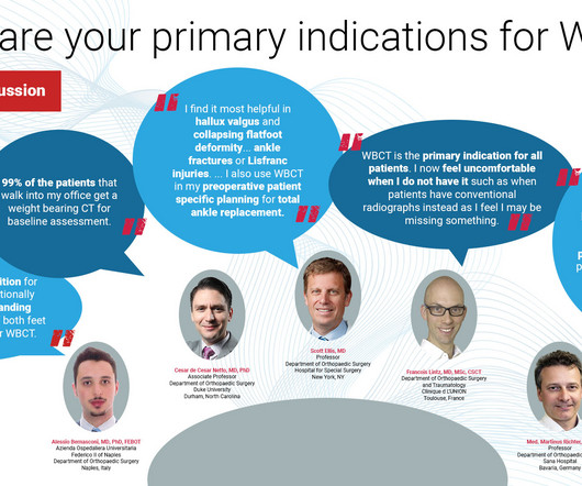
Alessio Bernasconi, MD, PhD, FEBOT Azienda Ospedaliera Universitaria Federico II of Naples Department of Orthopaedic Surgery Naples, Italy I think that every condition for which one would traditionally request a bilateral standing radiographic imaging of both feet is a good indication for WBCT. Even soft tissue issues (i.e.
With advance planning and an emphasis on learning, your facility can perform imaging studies with the same standard of care as a large children’s hospital. With advance planning and an emphasis on learning, your facility can perform imaging studies with the same standard of care as a large children’s hospital.
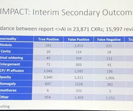
solutions are driving a significant improvement through early lung cancer detection within hospital systems. Averaged across all findings on chest radiographs.) [2]AIDE presented at ECR (European Congress of Radiology) 2024. million members. Lancet Digital Health. Published 2021. 2]AIDE study, Alfred Health. Data on file. [3]
Sites: Investigators recruited patients at 31 French emergency departments at university and nonuniversity hospitals Duration : June 1, 2009 to March 31, 2015. Funding: The Cardinal Health Laboratory provided Turkel thoracentesis kits to two hospitals Trial Registry: Clinicaltrials.gov: NCT01008228.
ClinicalTrials.gov: NCT02631759 Clinical Question : In adult patients aged 18 years or older presenting within 24 hours of a spontaneous ICH, does the administration of levetiracetam, compared to placebo, reduce the occurrence of clinical seizures? The paper presents interesting data. Lancet Neurology 2022; 21:781-91. PMID: 35963261.
The introduction of AI solutions, such as qXR-LN, presents a remarkable opportunity to cast a wider net to identify potentially malignant pulmonary nodules, thereby boosting the fight against lung cancer. Tailored for use in the incidental adult population, this innovative device is a game-changer in diagnostic technology.
Improved image processing looks that provide an even higher level of image quality with consistent presentation. Physicians have better diagnostic confidence when they can view radiographs in the manner most suitable to their preferences. Your radiographers need to work as efficiently as possible in order to keep pace.
Figure 2 A: AP view radiograph of right forearm. B: Lateral radiograph view of right forearm. An angulated fracture of the distal midshaft radius is also visualized, but there is also bowing of the ulna that is more appreciated on the lateral radiograph view. 8 Year Old Male With Trauma Due To A Fall. Xray of the Week Figure 1.
A) AP radiograph of Lisfranc Fracture Dislocation demonstrates the circled “fleck sign” or Lisfranc ligament avulsion fracture fragment. (B) C) The lateral radiograph notes with a circle, the dorsal sub dislocation of the metatarsal base. Trauma due to falling off a roof. Xray of the Week Figure 1. Trauma due to falling off a roof.
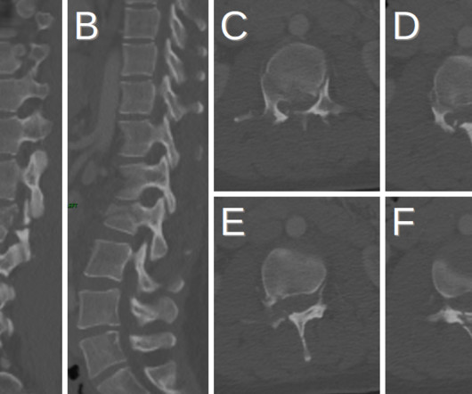
Fracture of the spinous process (blue arrows) is also present. Chance fractures are difficult to identify as they do not present with neurological deficits but can present with intra-abdominal injuries [1]. Plain radiograph shows empty vertebral body sign, which results from displacement of the spinous processes [3].
Patients with ectopia lentis typically present with visual acuity problems but can also present with eye pain if secondary to trauma [2]. Anterior axial lens subluxation, progressive myopia, and angle closure glaucoma: recognition and treatment of atypical presentation of ectopia lentis. Radiographics. 2006;10(4):345-350.
However, management continues to be debated and may lead to unnecessary hospitalization. PMID: 32622394 Clinical Question: Can ambulatory management of patients with primary spontaneous pneumothorax reduce the length of hospitalization? Often patients are otherwise young and healthy. Paper: Hallifax RJ et al. 2020;396(10243):39-49.
Additionally, the rate of diverticulitis appears to be increasing among both patients presenting to the Emergency Department and the population in general. Enrolled patients received follow-up assessments at 2, 7, 30, and 90 days after their initial presentation.
Adrian Brady, presidente de la Sociedad Europea de Radiología y radiólogo consultor del Hospital de la Universidad de Mercy en Cork, Irlanda. Es director médico del Hereditary Haemorrhagic Telangiectasia (HHT) (con sede en el Hospital de la Universidad de Mercy). Radiographics (2015);35:1668-1676.
It improves the Emergency department’s capacity & throughput whereas, in multispecialty tertiary hospitals, it works out as an alternate coverage for after-hours / vacation/weekends and plays important role in clearing the department’s stack. Our team stands up to the statement on quality as briefed in Radiographics, 2011.
Like pyelonephritis, patients with EPN often present with fever, abdominal or flank pain, and costovertebral angle tenderness. KUB indicates kidneys, ureter, and bladder (plain abdominal radiograph); CT, computed tomography; and PCD, percutaneous catheter drainage. RadioGraphics. 2000;160(6):797-805. doi:10.1001/archinte.160.6.797
These applications, cited by Jesbon, include diagnostic functions such as detecting pneumonia on chest radiographs or grading liver tumors, as well as repetitive tasks like breast or lung nodule detection. AI, with its ability to enhance efficiency and reduce costs, will be critical in helping these departments navigate these challenges.
Photoprint from radiograph by W.K. Within weeks of his announcement hospitals world-wide had taken the initiative to open up X-ray rooms, which gave rise to the first radiology departments. (3) Portrait of Sir Godfrey Hounsfield (1919-2004) The first clinical CT scan: Atkinson Morley's Hospital, October 1971 Credit: impactscan.org.
Cesar de Cesar Netto, MD, PhD Associate Professor Duke University Durham, North Carolina Dr. Scott Ellis, MD Professor Hospital for Special Surgery New York, NY Hallux Valgus : I find it very helpful to assess the amount of first metatarsal pronation present as part of the hallux valgus deformity along with sesamoid arthritis and position.
Thin-section axial CT scans followed by multiplanar reformations can visualize ruptured globe contour, which can present as a “mushroom” (Figs. The patient’s presenting visual acuity is a major determinant of their post-surgical visual acuity. Radiographics. 1-2) or “flat tire” shape. Clin Ophthalmol. 2013;7:461-465.
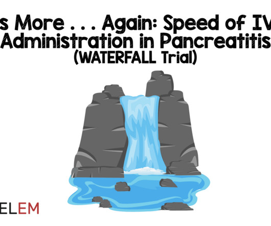
Population: Adult patients > 18 years of age diagnosed with acute pancreatitis based on the Revised Atlanta Classification (requires 2 of 3: typical abdominal pain, serum amylase or lipase level higher than 3 times the upper limit of normal or signs of acute pancreatitis on imaging) that presented within 24 hours of pain onset.
The synthetic control arm was obtained retrospectively from one of three acute care hospitals in the Hartford Healthcare network between 12/1/2016 and 8/30/2020. POPULATION Inclusions: Age ≥ 18 years of age Radiographically confirmed acute spontaneous or traumatic intracranial hemorrhage (i.e. vs. 24.2%). vs. 24.2%). vs. 14.7%).
We organize all of the trending information in your field so you don't have to. Join 5,000 users and stay up to date on the latest articles your peers are reading.
You know about us, now we want to get to know you!
Let's personalize your content
Let's get even more personalized
We recognize your account from another site in our network, please click 'Send Email' below to continue with verifying your account and setting a password.


Let's personalize your content