This site uses cookies to improve your experience. To help us insure we adhere to various privacy regulations, please select your country/region of residence. If you do not select a country, we will assume you are from the United States. Select your Cookie Settings or view our Privacy Policy and Terms of Use.
Cookie Settings
Cookies and similar technologies are used on this website for proper function of the website, for tracking performance analytics and for marketing purposes. We and some of our third-party providers may use cookie data for various purposes. Please review the cookie settings below and choose your preference.
Used for the proper function of the website
Used for monitoring website traffic and interactions
Cookie Settings
Cookies and similar technologies are used on this website for proper function of the website, for tracking performance analytics and for marketing purposes. We and some of our third-party providers may use cookie data for various purposes. Please review the cookie settings below and choose your preference.
Strictly Necessary: Used for the proper function of the website
Performance/Analytics: Used for monitoring website traffic and interactions
Some of my radiological heroes would report a staggering 30,000 to 40,000 radiographs a year. Some even [startled gasp] gave up reporting plain radiographs. But the pressure kept building, and the number of CT and MRI scans grew by 20% annually in my hospital. I still don’t know how they did it. This was all delegated.
"Practical AI implementation will require objective onsite performance evaluation, institutional information technology infrastructure integration, and postdeployment monitoring," wrote a team led by Eui Jin Hwang, MD, PhD, of Seoul National University Hospital in South Korea.
. | S1-SSCH01-5 | E451A This scientific paper may increase overall confidence in the potential of using multimodal AI for tuberculosis (TB) detection, and potentially autonomous reporting, on chest radiographs in certain clinical settings.
The finding is from a study conducted by a group at the University Hospital Jena in Eisenberg, Germany, that explored whether hip x-rays could be reliable indicators for bone mineral density (BMD) in male and female patients prior to THA procedures. “An
The finding by researchers in Copenhagen, Denmark, suggests that AI could eventually help streamline high-volume radiology workflows by handling some of the more “tedious parts of the work,” lead author Louis Plesner, MD, of Herlev and Gentofte Hospital in Denmark told AuntMinnie.com.
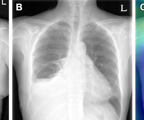
Radiologists routinely compare the current and previous chest radiographs during interpretation to enhance the sensitivity for change detection and provide information for differential diagnosis. Example of triage of no change in a pair of chest radiographs in the emergency department. (A) Image courtesy of Radiology.
. | W3-SSMK08-4 | Room E450A A deep learning-based framework for automated screening of osteoporosis on lumbar spine plain radiographs shows potential as another way to opportunistically make use of imaging studies performed for other indications, according to this presentation.
The deep learning (DL) model may guide clinical decision-making in the management of patients with CAP by identifying high-risk patients who warrant hospitalization and intensive treatment,” concluded first author Eui Jin Hwang, MD, PhD, from the department of radiology at Seoul National University College of Medicine in Korea. Hwang et al.
Gleamer) increased sensitivity for detecting all abnormalities on x-rays for all readers (thoracic radiologists, general radiologists, and radiology residents), according to a group of Gleamer consultants and clinicians at Cochin Hospital in Paris. In a retrospective study, a commercially available algorithm (ChestView, v.
Charlene Liew, MD, director of cardiothoracic imaging at Changi General Hospital in Singapore, outlined the current state of AI in radiology and where it is going in a presentation called "AI in Radiology: The Past Informs the Future."
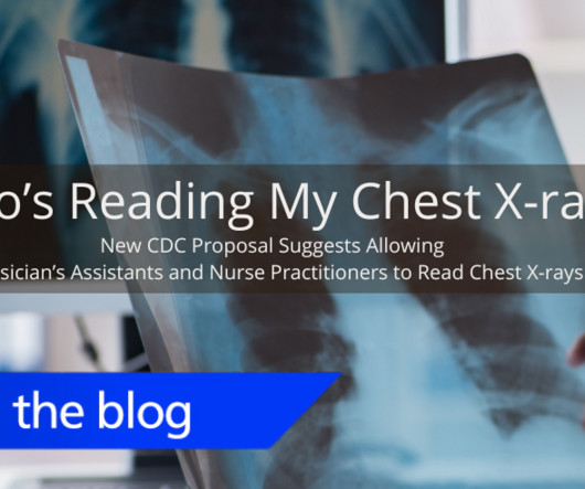
This program trains and certifies physicians to read chest radiographs for workers participating in health surveillance programs. Radiologists complete over 20,000 hours of clinical experience before being certified to interpret radiographic images. Compared to only 500 hours of clinical experience for NPs and 2000 hours for PAs.
I'm a radiographer,' " Stewart recalled. AuntMinnie.com · Minnies 2024 - Most Effective Radiologic Sciences Educator Runner-up: Colleen Dempsey, EdD, Thomas Jefferson University, Philadelphia, PA Most Effective Radiology Administrator/Manager Minnies 2024 Winner: Dmitry Beyder, CNMT, Barnes-Jewish Hospital / Washington University, St.
“This method plays an important role in overcoming the barriers to its clinical implementation, including the need for trained experts and the time-consuming process of manual contouring,” noted lead author Noriaki Wada, MD, of Brigham and Women’s Hospital in Boston. The subject is a 77-year-old male with VC of 1.68 L and FEV1 of 0.68
The first scans have been performed in the Olympic imaging polyclinic ahead of Friday's opening ceremony, and the 68-strong squad of radiologists and radiographers are primed and ready for action, according to musculoskeletal (MSK) expert Jérôme Renoux, MD. Bring it on!
A team led by Sofyan Jankowski, MD, of Lausanne University Hospital in Switzerland found no statistically significant difference between ChatGPT’s answers and those posted on radiology institutional websites. and Europe to evaluate both sets of answers, blinded to the source.
They then trained the algorithm on a data set of 47,150 x-rays (23,151 patients) and validated it on an external data set of 2,914 radiographs (1,515 patients). The model achieved 79% accuracy, 96.6% sensitivity, and 34.1% specificity in predicting T-scores ≤ -1.0 normal) from the x-rays, while T-score predictions ≤ -2.5 accuracy, 77.1%
The Karolinska hospital group has recently established an MR safety team involving staff at Solna and Huddinge. We have registered the accident with the authorities and started an internal investigation, but we will wait for the report on what caused the accident before we take any further steps.
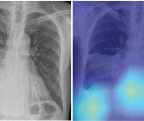
Presenting the research on November 28 at RSNA 2023, Jan Rudolph, MD, from the department of radiology at University Hospital LMU Munich said nonradiologists can significantly benefit from AI assistance in emergency-related chest x-ray analysis. “If
A team led by Yin Ting Chiu, PhD, from the Hong Kong Children’s Hospital discussed key factors identified by the hospital’s MR team for effectively performing supplementary MRI scans on children ages three to seven without sedation.
It's a question researchers at University College London Hospitals NHS Foundation Trust and Canterbury Christ Church University have asked as part of their prospective, randomized, multisite trial currently open and underway in the U.K. How does immediate AI-enabled patient triage on chest CT impact the lung cancer pathway?
A team led by Junqi Han, MD, from the Affiliated Hospital of Qingdao University in China found that its model combining data from mammography images, ultrasound images, and other characteristics performed well in predicting disease-free survival of breast cancer.
He presented the abstract on behalf of first author Ken Herrmann, MD, chair of nuclear medicine at the University Hospital Essen in Essen, Germany. Radiographic progression-free survival (rPFS) was the primary endpoint of the study, while overall survival was the key secondary endpoint.
Louis School of Medicine and completed his residency and a neuroradiology fellowship at Massachusetts General Hospital in Boston. Dempsey began working in radiography at Mercy Fitzgerald Hospital in Darby, Pennsylvania. She continued working at the hospital for 12 years while growing in her role as an educator. Dmitry Beyder.
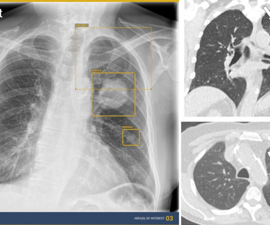
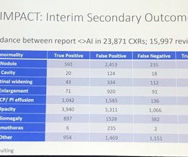
This competition demonstrated the value of AI in detecting and localizing many pathologies in chest radiographs by simulating the real work situations of radiologists,” the group wrote. The study was published February 8 in the Journal of Imaging Informatics in Medicine.
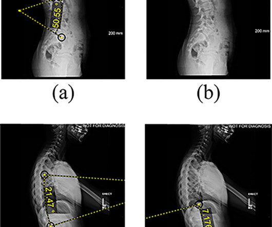
The researchers included data from 91 patients with adolescent idiopathic scoliosis who underwent AVBT surgery at the Shriners Hospitals for Children in Philadelphia. Features extracted from lateral radiographs. (a) Hence, the group developed a machine-learning-based algorithm that could potentially fill this gap.
The green project conducted by radiographers at the European Institute of Oncology in Milan and nearby Hospital of Legnano saved an estimated 12,000 euros ($13,000), said Andrea Masperi, who presented the details. during indirect radiology department activities, according to findings of a pilot study presented March 1 at ECR 2024.
Whenever bilateral standing radiographs would have been needed, a WBCT was performed instead. The reality is that if insurance was not an issue, I would not perform conventional radiographic imaging anymore. It is usually when I am trying to prove instability and the need for surgery.
A group at Mount Sinai Hospital developed a “pipeline” of convolutional neural networks (CNNs) to analyze lung areas in DDR image sequences from patients. a) Raw example of a dynamic digital radiograph. (b) The model performed well enough to act as a surrogate to standard pulmonary function tests, they found.
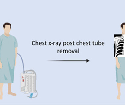
Ordering x-rays after removing chest tubes from lung surgery patients does not lead to meaningful change in patient management and prolongs hospital stays, according to a study published July 26 in the Journal of Thoracic and Cardiovascular Surgery. No patient underwent any procedure following the post-chest tube removal x-ray. days vs. 2.3
In an open forum, Yi Xiang Tay, of Singapore University Hospital's radiography and diagnostic imaging department, shared his team's research. It was the endnote of a series of sessions focused on optimizing radiology services.
Hospitals must develop a "safety first" culture for emergency department MRI, which should include tight access control, mandatory staff training, and vigorous screening, according to Mandel. There must be sufficiently trained radiology staff to avoid [injuring] people, [and that's why] MRI radiographers must be in charge in all MRI areas."
Repeating imaging exams increases the workload of your radiographers who are already stretched too thin; increases the exposure of the affected patients; and contributes to patients’ reduced confidence and satisfaction with your imaging department. The Audio Assist makes it easier for radiographers to hear the patients.
ARRS President’s Award for “Multi-task Ensemble Deep Learning for Differential Diagnosis of Pneumonia and Pulmonary Edema on Chest Radiograph.” Haver, MD, Massachusetts General Hospital in Boston. Ahmed Taher, MD, University of Texas Health Science Center at Houston. Versus GPT-4 in Assigning BI-RADS Final Assessment Categories.”
Benzaia, who is known for his on-screen medical professional roles in shows such as "General Hospital," "How to Get Away with Murder," "Law & Order," and "The Blacklist," has also worked as a radiographer and CT technologist.
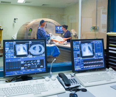
Additionally, all table movements can be controlled by the radiographer from the touch screen, enabling easy setup of either fluoroscopy or radiography settings. A camera integrated into the collimators allows the radiographer to position the patient directly from the console without using fluoroscopy.
When the radiology department at Cork University Hospital in the south coast of Ireland took the lead in developing an autism-friendly patient experience, they observed that it took less time to complete imaging studies for children with autism, according to a February 28 session at ECR 2024.
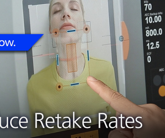
The white paper evaluates the capabilities of Canon Intelligent NR and its deep learning neural network (DLNN) software in relation to standard and decreased dose pediatric digital radiographs at Dayton Children’s Hospital, Dayton, Ohio. 26 –29, 2023 in North Hall, Level 3, booth #7913.
Earlier this spring, Carestream’s Digital Marketing Manager, John Crowther, had the opportunity to travel to Shriner’s Children’s Hospital in St. Aligned Goals Carestream’s solutions help deliver an enhanced experience for both patient and staff at Shriner’s Children’s Hospital in St. Here is his experience.
The first female editor in chief of RSNA’s peer-reviewed RadioGraphics journal since 2021, Menias splits her time between Mayo Clinic, where she’s a specialist in abdominal imaging, and the journal, where she is achieving her first goals of expanding RadioGraphics as a learning platform.
solutions are driving a significant improvement through early lung cancer detection within hospital systems. Averaged across all findings on chest radiographs.) [2]AIDE million members. Radiologists using Harrison.ai's technology have seen an over 45% increase in diagnostic accuracy1. Lancet Digital Health. Published 2021.
Reading Time: 10 minutes read By Henry Williams, Carestream Area Vice President, Sales Western Nowadays, with hospital budgetary restrictions at the forefront of the purchasing decision making process, it seems like the X-Ray market, like everything else, is not immune to the current state of the economy. Who is Making the Purchases?
Key Points: Currently plain radiographs are the standard method in diagnosing syndesmotic ankle injuries even though the distal tibiofibular joint cannot be assessed due to superposition of the osseous structures in the foot. Dr. Peiffer et.
A team of 32 radiologists and 36 radiographers are limbering up to work at the summer Olympics, which begins July 25. If they need more acute care, they will be referred to the local hospital,” he explained. They are likely to perform more than 1,800 scans on injured athletes.
The enduring shortage is affecting staffing for radiographers and radiologists; and all imaging modalities. Travel positions offer better pay while the increasing number of urgent care and free-standing emergency centers offer an environment that is more attractive and less stressful to many candidates than a hospital.
We organize all of the trending information in your field so you don't have to. Join 5,000 users and stay up to date on the latest articles your peers are reading.
You know about us, now we want to get to know you!
Let's personalize your content
Let's get even more personalized
We recognize your account from another site in our network, please click 'Send Email' below to continue with verifying your account and setting a password.


Let's personalize your content