This site uses cookies to improve your experience. To help us insure we adhere to various privacy regulations, please select your country/region of residence. If you do not select a country, we will assume you are from the United States. Select your Cookie Settings or view our Privacy Policy and Terms of Use.
Cookie Settings
Cookies and similar technologies are used on this website for proper function of the website, for tracking performance analytics and for marketing purposes. We and some of our third-party providers may use cookie data for various purposes. Please review the cookie settings below and choose your preference.
Used for the proper function of the website
Used for monitoring website traffic and interactions
Cookie Settings
Cookies and similar technologies are used on this website for proper function of the website, for tracking performance analytics and for marketing purposes. We and some of our third-party providers may use cookie data for various purposes. Please review the cookie settings below and choose your preference.
Strictly Necessary: Used for the proper function of the website
Performance/Analytics: Used for monitoring website traffic and interactions
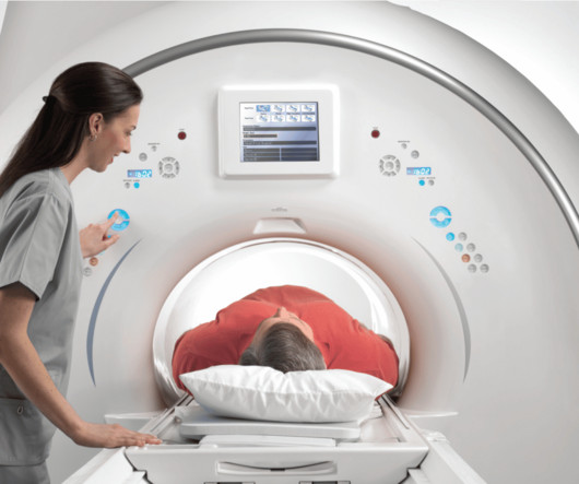
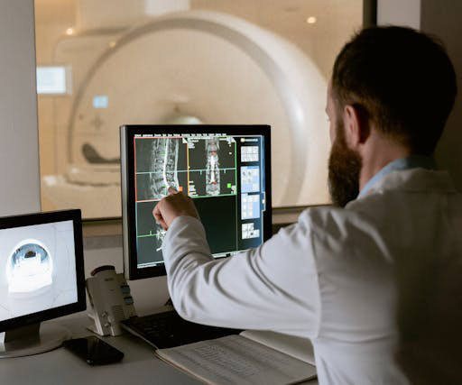
Imageinterpretation can be one major source of variability, with some radiation oncologists forced to read MR, CT and other exams during the RT planning process.
However, mpMRI misses about 10% of cases, typically in patients with lower-grade disease and in patients with cribriform pattern disease, a subtype much more likely to recur after surgery or radiation therapy, he noted. Interpretation accuracy is compared with biopsy results.
Many radiation oncologists are not formerly trained in imaginginterpretation, and radiologists’ collaborative participation in care planning can help to catch errors, experts wrote recently.
The college represents thousands of radiologists, radiation oncologists, nuclear medicine physicians, and physicists. The institution is known as “the voice of radiology,” as they are at the forefront of innovation and therefore responsible for regulation and legislation around the field. Today, the College has 54 chapters.
This includes more streamlined processing, structured reporting, fast and high-quality contouring, advanced image fusion, dosimetry capabilities, easier IT integration, and remote access – helping to boost healthcare productivity and promote personalized patient care. There is also special focus on radiation therapy and Theranostics.
PET imaging with POSLUMA reveals clinical information crucial to decision-making for men with prostate cancer, and we are excited to share further information with the radiation oncology community at ASTRO 2023,” said David E. Jani, MD, FASTRO, Department of Radiation Oncology, Winship Cancer Institute of Emory University, Atlanta, Ga.,
A negative image does not rule out the presence of prostate cancer and a positive image does not confirm the presence of prostate cancer. POSLUMA use contributes to a patient’s overall long-term cumulative radiation exposure. Long-term cumulative radiation exposure is associated with an increased risk for cancer.
Chapter 5: Multidetector CT – Advancements in Image Reconstruction: Discover how advancements like multidetector CT have transformed the image reconstruction process, improving image quality and efficiency.
Teleradiology-India Introduction: While the primary role of teleradiology is in diagnostics, its impact extends beyond imageinterpretation. Discuss how detailed imaging data contributes to radiation therapy planning, enabling oncologists to deliver targeted and precise treatments.
Immediate Diagnostic Insights: Discuss the critical role of Night Hawk Radiology in offering immediate imageinterpretations, particularly in emergency and critical care situations, and how this precision impacts patient care. Section 2: Precision in the Dark 2.1
Innovations in X-ray Technology: Recent advancements in X-ray technology include low-dose imaging techniques to reduce radiation exposure, dual-energy X-ray absorptiometry (DXA) for bone density assessment, and cone-beam CT for 3D dental imaging. The Future of X-ray Technology: The future of X-ray technology is bright.
It is non-invasive and can determine the effectiveness of radiation treatments and other important information, such as cell density and microstructure of the tissue. In addition, the combination of PET/MRI imaging is proving to be even more powerful than MRI alone.
Radiologist’s Expertise: The Art of ImageInterpretation: Emphasize the expertise of radiologists in the interpretation of medical images. Challenges in Radiological Imaging: Navigating Complexity: Address challenges faced in radiological imaging.
“Our study showed evidence of hallucinatory responses when interpretingimage findings,” Dr. Klochko said. “We We noted an alarming tendency for the model to provide correct diagnoses based on incorrect imageinterpretations, which could have significant clinical implications.” Radiology is edited by Linda Moy, M.D.,
One of his tweets: #RGchat T1: the ABR certification exam is intended to test knowledge as it relates to competence, and critical thinking as it relates to imageinterpretation. I called it “ The ABR Defines the Intent of the Core Exam ,” and I think it’s worth reading for our discussion here.
We are pleased to present these results from the LIGHTHOUSE study to the radiation oncology community at ASTRO,” said David E. A negative image does not rule out the presence of prostate cancer and a positive image does not confirm the presence of prostate cancer. Gauden, D.Phil.
This approach enables radiologists to focus on imageinterpretation rather than transcribing numbers, thereby reducing dictation time and minimizing errors. demonstrated the effectiveness of checklists in reducing imaging costs for ultrasound screening of suspected appendicitis in pediatric patients [ 8 ].
This approach enables radiologists to focus on imageinterpretation rather than transcribing numbers, thereby reducing dictation time and minimizing errors. demonstrated the effectiveness of checklists in reducing imaging costs for ultrasound screening of suspected appendicitis in pediatric patients [ 8 ].
tesla MRI AI body composition analysis Cardiac PET Cryo/thermoablation CT colonography Genicular artery embolization Hyperpolarized xenon-129 MRI PET/MRI Photon-counting CT Radiomics Theranostics Whole-body MRI screening Image of the Year 3D PET/MR image. Workplace violence in medical radiation science: A systematic review.
3) The British Röntgen Society (the first radiology society) was founded in 1897, and many further studies on X-ray usage and the effects of radiation were performed over the following years. (3) Computerized follow-up of discrepancies in imagedinterpretation between emergency and radiology departments. Prevedello.
We organize all of the trending information in your field so you don't have to. Join 5,000 users and stay up to date on the latest articles your peers are reading.
You know about us, now we want to get to know you!
Let's personalize your content
Let's get even more personalized
We recognize your account from another site in our network, please click 'Send Email' below to continue with verifying your account and setting a password.


Let's personalize your content