This site uses cookies to improve your experience. To help us insure we adhere to various privacy regulations, please select your country/region of residence. If you do not select a country, we will assume you are from the United States. Select your Cookie Settings or view our Privacy Policy and Terms of Use.
Cookie Settings
Cookies and similar technologies are used on this website for proper function of the website, for tracking performance analytics and for marketing purposes. We and some of our third-party providers may use cookie data for various purposes. Please review the cookie settings below and choose your preference.
Used for the proper function of the website
Used for monitoring website traffic and interactions
Cookie Settings
Cookies and similar technologies are used on this website for proper function of the website, for tracking performance analytics and for marketing purposes. We and some of our third-party providers may use cookie data for various purposes. Please review the cookie settings below and choose your preference.
Strictly Necessary: Used for the proper function of the website
Performance/Analytics: Used for monitoring website traffic and interactions
Women’s imaging at this year’s RSNA reflects momentous changes in trends regarding personalized, targeted healthcare in 2024 for women’s health. Research efforts toward supplemental breast imaging have ramped up in recent years and in Chicago, attendees can see results from these imaging modalities being put to the test.
Imaging AI tools and algorithms continue to be rapidly developed and deployed into clinics, but experts say theres an elephant in the room that still needs to be addressed: reimbursement. Specifically, AI supporters are focusing on the lack of current procedural terminology (CPT) codes representing imaging services performed with AI.
When it comes to medical imaging, radiology is what most often comes to mind, and for good reason. A large percentage of medical imaging created by most hospitals tends to come from the radiology department. In most cases, medical images and scans are placed into an electronic medical record (EMR) as a link. In the U.S.
To better predict breast density, some researchers have turned to radiomics, where quantitative parameters are created from sets of images. They investigated which breast images could best predict breast density in these models. The team extracted a total of 123 imaging features from the 10 regions of interest of 96 women.
In this guide, we’ll take you through a step-by-step process to transform your radiology center into a high-performance hub of medical imaging. Patient-centric scheduling can only be achieved through optimized radiology workflows, effective communications between staff and physicians, and, of course, through specialized schedulers.
Independent imaging centers are experiencing increased competition for CT patients, according to the newly published IMV 2024 CT Market Outlook Report. Hospital-based sites are more likely to be experiencing this shift compared to independent imaging centers. Independent imaging centers 10.9 hours on weekdays and 17.9
A Swiss research team has developed and tested an AI model that automatically segments anatomic structures on MR images independent of sequence, according to a study published February 18 in Radiology. Images and caption courtesy of the RSNA. The team then assessed the model's performance using Dice scoring.
The way physicians identify illness is changing due to advances in medical imaging, which make early diagnosis quicker, more precise, and less invasive. As one of El Pasos top radiology centers, Professional Radiology is dedicated to offering cutting-edge imaging services that aid in the early detection of illnesses.
Food and Drug Administration (FDA) has issued draft guidance providing recommendations to drug companies on the design of clinical trials for optical imaging drugs used with imaging devices during medical procedures. The FDA is taking comments on the guidance through April 8, 2025.
While academic medical center radiology departments are expanding significantly and hospitals are adapting to health system consolidation trends, demand for innovative imaging informatics remains strong among operations and physician teams. billion healthcare facility. Another, a new hospital complex is underway at University of Utah Health.
Using a deep-learning model with lung CT imaging shows promise for identifying and segmenting tumors, researchers have reported. To address the problem, Kashyap and colleagues developed and tested a 3D U-Net-based, image-multiresolution ensemble deep-learning model for identifying and segmenting lung tumors on CT scans.
Finding the right enterprise imaging (EI) system is critical for radiology practices and hospitals that need to expand and scale their multi-specialty image management and reading capacity.
Implementing a clinical decision support system (CDSS) had little effect on reducing inappropriate imaging orders by doctors in university hospitals, according to a study published February 10 in JAMA. The primary outcome measure was the proportion of inappropriate imaging requests made per department before and after implementation.
The use of AI in thoracic imaging has begun to demonstrate "cumulative evidence of effectiveness," but more testing and research are needed to determine its feasibility for this application, according to a commentary published February 25 in Radiology. van Beek, MD, of the University of Edinburgh in the U.K. in an accompanying editorial.
Radiologist Jelle Barentsz, MD, PhD, of Radboud University in the Netherlands, emphasized that poor-quality MRI images are leading to prostate cancer overdiagnosis. We need to have a quality measurement of the imaging," he explained. "We We developed PI-QUAL which is, I think, the first MRI standard looking at image quality.
Radon Medical Imaging has purchased Alpha Imaging, a large imaging equipment distributor and services provider based in Willoughby, OH. Alpha Imaging’s portfolio includes cardiovascular and interventional imaging labs and digital radiographic and fluoroscopic systems. Terms of the transaction were not disclosed.
Artificial intelligence has the potential to improve the analysis of medical image data. This is the result of AutoPET, an international competition in medical image analysis. For example, algorithms based on deep learning can determine the location and size of tumors.
There's plenty of research and technologies on display at RSNA 2024 when it comes to breast imaging. She also talked about what breast imaging facilities can expect with the U.S. She also talked about what breast imaging facilities can expect with the U.S. Just ask Amy Patel, MD, from Liberty Hospital in Missouri.
The software has been primarily disguised as Philips DICOM MediaViewerLauncher.exea trusted program that enables patients to view their medical imaging on their own personal servers.
celebrates this year's Breast Cancer Awareness Month, radiology experts and advocates reflect on the big year that breast imaging has had. The mandate requires imaging facilities to disclose breast density information to patients receiving breast cancer screening. As the U.S. From the U.S.
Imaging AI software developer HOPPR has added Woojin Kim, MD, and William Boonn, MD, to its executive lineup. A musculoskeletal radiologist, imaging informaticist, and entrepreneur, Kim comes to HOPPR after serving most recently as chief medical information officer (CMIO) at Rad AI.
Komen applauded the passage of breast imaging legislation in Virginia that the organization said eliminates barriers to essential care. SB 1436 will allow people with state-regulated health plans to receive diagnostic and supplemental imaging without high patient cost sharing, Komen said.
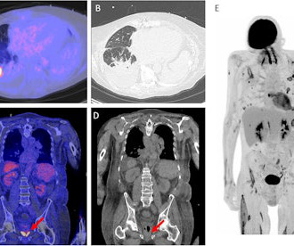
There are several conceivable advantages of F-18 FDG PET/CT over conventional imaging. Image A and B showing an example of the distinctive capability of the F-18 FDG PET/CT. A shows a fusion image of F-18 FDG PET/CT and low-dose CT and B an high resolution chest CT of a patient with an Aspergillus infection. DEED, Attribution 4.0
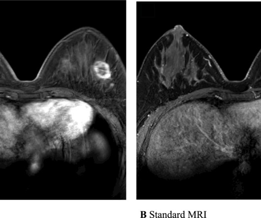
Features used for radiomics models can provide systematic analysis of images by overcoming the limitations of subjective analysis and dependence on the experience of radiologists interpreting MR images. From there, the researchers extracted 1,618 radiomic features and four kinetic features from ultrafast and standard MR images.
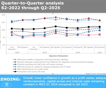
Radiology administrators express very high confidence that imaging will grow as a profit center in coming months, according to The MarkeTech Group's Medical Imaging Confidence Index (MICI) report for the second quarter of 2025. This is a national crisis that will only get worse."
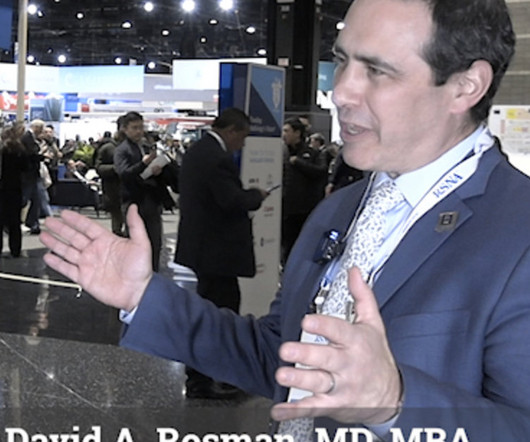
Rosman, MD, MBA, deputy chief, radiology enterprise service, Mass General Brigham, explains how moving imaging outside of hospitals could save billions of dollars for U.S. healthcare.
Agfa HealthCare and enterprise imaging marketplace platform firm Carpl.ai RUBEE for AI into Agfa HealthCare’s enterprise imaging system. Agfa stated that RUBEE can integrate directly into Agfa HealthCare enterprise imaging systems and is designed to work with already trusted tools.
Conversely, prostate-specific membrane antigen (PSMA) PET is a molecular imaging approach that uses radiotracers to detect these types of cancer based on the expression of PSMA protein by the cancer cells. Hybrid PET/MRI scanners were introduced about 15 years ago to leverage the advantages of both methods.
Those proposing breast imaging-related measures so far in 2025 have included Connecticut, Florida, Hawaii, Indiana, Missouri, New York, South Carolina, Utah and Virginia, ACR reports.
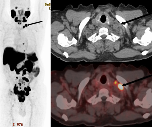
PET/CT scans with an experimental prostate-specific membrane antigen (PSMA) imaging agent can identify supraclavicular nodal metastasis in newly diagnosed prostate cancer patients, researchers have reported. Image and caption available for republishing under Creative Commons license (CC BY 4.0 DEED, Attribution 4.0
The Los Angeles-based imaging center operator recently inked a partnership with a Florida OB-GYN group and plans to "aggressively" pursue similar opportunities.
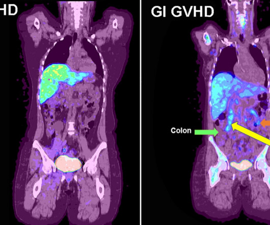
F-18 fluorothymidine (FLT) PET can identify early acute gastrointestinal graft versus host disease (GVHD) after patients undergo bone marrow transplants, according to a study published December 13 in Radiology: Imaging Cancer. F-18 FLT-PET imaging was performed on day 28. F-18 FLT-PET imaging of gastrointestinal GVHD.
Behind him is an image created by one such generative AI model, which illustrates humans working alongside AI. In radiology, the technologys performance has been assessed in response generation to patients, taking board exams, and image interpretation assistance. Kohli used generative AI to create some of his presentation slides.
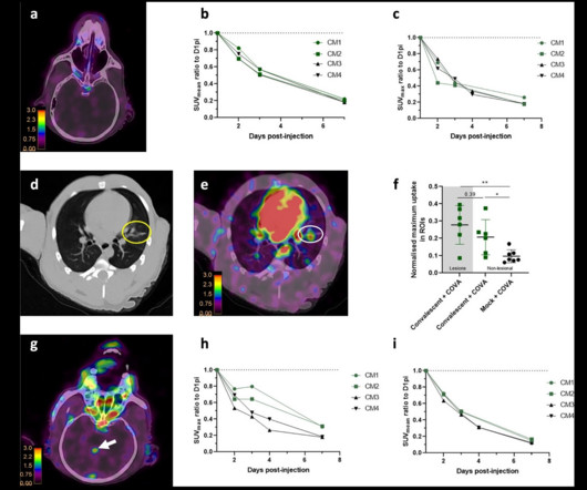
Researchers in France have developed a PET imaging approach for longitudinal tracking of the virus that causes COVID-19. Image and caption available for republishing under Creative Commons license (CC BY 4.0 The group detected Zr-89 COVA1-27-DFO uptake by the lungs of one convalescent animal, along with CT ground-glass opacity.
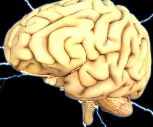
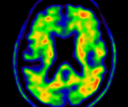
Researchers examined the brains of bilingual and monolingual people using fMRI, only to find improved communication between isolated regions in those who learn a second language at a young age.
Payers have reportedly use artificial intelligence algorithms to deny routine requests for PET imaging, instead steering patients to CCTA in some cases.
Excess imaging translates to "significant"carbon dioxide (CO2) emissions, according to a study published March 28 in the Journal of the American College of Radiology. Overuse of imaging is a strain on our healthcare system and radiology workforce," said senior author Michael Atalay, MD, PhD, of Brown University in Providence, RI.
Amy Klobuchar, D-Minn., and Roger Marshall, MD, R-Kan., introduced the companion version of the Find It Early Act over a year after the House bill was proposed.
The new appropriate use criteria define 17 specific clinical scenarios, guiding providers on situations when amyloid or tau imaging are and are not appropriate.
Ga-68 FZ-NR-1 PET/CT images and F-18 FDG PET/CT and PET/MRI images in representative nectin-4-positive TNBC patients. (A) A) PET/CT images and nectin-4 immunohistochemistry (IHC) staining of patient 1. B) PET/CT images and nectin-4 IHC staining of patient 2. Image courtesy of the Journal of Nuclear Medicine.
We organize all of the trending information in your field so you don't have to. Join 5,000 users and stay up to date on the latest articles your peers are reading.
You know about us, now we want to get to know you!
Let's personalize your content
Let's get even more personalized
We recognize your account from another site in our network, please click 'Send Email' below to continue with verifying your account and setting a password.


Let's personalize your content