This site uses cookies to improve your experience. To help us insure we adhere to various privacy regulations, please select your country/region of residence. If you do not select a country, we will assume you are from the United States. Select your Cookie Settings or view our Privacy Policy and Terms of Use.
Cookie Settings
Cookies and similar technologies are used on this website for proper function of the website, for tracking performance analytics and for marketing purposes. We and some of our third-party providers may use cookie data for various purposes. Please review the cookie settings below and choose your preference.
Used for the proper function of the website
Used for monitoring website traffic and interactions
Cookie Settings
Cookies and similar technologies are used on this website for proper function of the website, for tracking performance analytics and for marketing purposes. We and some of our third-party providers may use cookie data for various purposes. Please review the cookie settings below and choose your preference.
Strictly Necessary: Used for the proper function of the website
Performance/Analytics: Used for monitoring website traffic and interactions
AI software based on deep-learning algorithms is showing promise, however, for helping to improve specificity in screening mammography and other breast imagingmodalities. million mammograms were performed in the U.S. Although mammogram is the most widely used screening modality, a known problem is that 9.5%
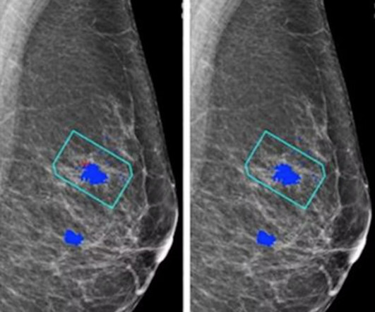
Postoperative MRI surveillance appears to lower the odds of advanced second breast cancer in women with a personal history of the disease, researchers have reported. "In Images in a 40-year-old woman who underwent breast-conserving surgery for left breast cancer and a surveillance breast MRI examination 25 months after surgery. (A)
Low-dose positron emission mammography (PEM) can detect invasive breast cancer in a feasible manner, according to research published February 9 in Radiology: Imaging Cancer. This study underscores the potential of this low-dose PEM system as a promising imaging tool in breast cancer diagnosis,” the Freitas team wrote.
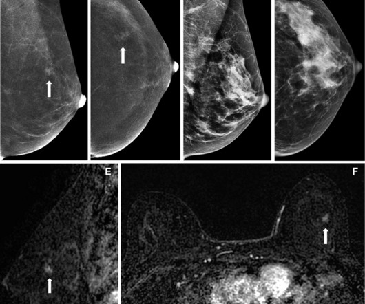
A team led by Julie Hamzah, MBBS, from Singapore General Hospital, found that symptomatic first breast cancers, dense breasts, and the presence of trabecular thickening on mammography are tied to mammogram detection failure of ipsilateral second breast cancers.
Food and Drug Administration (FDA) has said CT is still the preferred imagingmodality for patients with medical devices. CT is safer than magnetic resonance imaging (MRI) for people with devices of unknown MRI safety status." Read the FDA's full guidance here.
A team led by Joao Horvat, MD, from the Memorial Sloan Kettering Cancer Center in New York found that CEM depicted 90% of breast cancers compared with 10% on low-energy mammograms alone and 50% on low-energymammogramswith whole-breast ultrasound. Horvat and co-authors investigated whether the same trend goes for CEM.
For dense-breasted patients requiring supplemental imaging, MRI remains a valuable option that is not limited by breast density and is shown to be more sensitive than mammography at finding breast cancer. Ultrasound rounds out the radiologist’s toolkit for supplemental imaging of women with dense breasts.
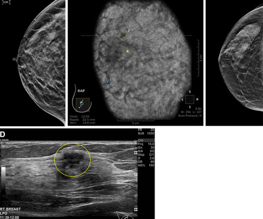
Since almost half of the screening population has dense breasts, many of these patients require additional breast imaging, often with MRI , after mammography. PEM displayed comparable performance to MRI, identifying 24 of the 25 invasive cancers (96%). Its false positive rate was only 16%, compared with 62% for MRI.
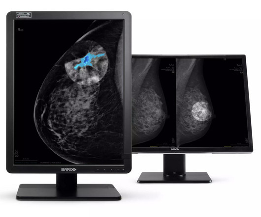
Marissa Fayer concludes: “Technologies like DL Precise offer new hope for the millions of people whose dense breast tissue currently masks cancer on mammograms.” For more information: www.barco.com Find more RSNA23 conference coverage here Monday, November 27, 2023 - 14:06 If you enjoy this content, please share it with a colleague
These findings can help breast imagers estimate the expected outcomes of supplemental ultrasound screening according to a woman’s risk level and assist in determining which women with dense breasts may be good candidates for supplemental ultrasound screening after a negative mammogram,” Sprague told AuntMinnie.com.
In the 1960s and 1970s, scientific research was published about the diffusion, relaxation and chemical exchange of water intracellularly, eventually leading to Magnetic resonance imaging (MRI). (6) 9) This image nearly took him 5 hours to acquire. Their work gave rise to the modern MRI scanners we use today.
We organize all of the trending information in your field so you don't have to. Join 5,000 users and stay up to date on the latest articles your peers are reading.
You know about us, now we want to get to know you!
Let's personalize your content
Let's get even more personalized
We recognize your account from another site in our network, please click 'Send Email' below to continue with verifying your account and setting a password.


Let's personalize your content