This site uses cookies to improve your experience. To help us insure we adhere to various privacy regulations, please select your country/region of residence. If you do not select a country, we will assume you are from the United States. Select your Cookie Settings or view our Privacy Policy and Terms of Use.
Cookie Settings
Cookies and similar technologies are used on this website for proper function of the website, for tracking performance analytics and for marketing purposes. We and some of our third-party providers may use cookie data for various purposes. Please review the cookie settings below and choose your preference.
Used for the proper function of the website
Used for monitoring website traffic and interactions
Cookie Settings
Cookies and similar technologies are used on this website for proper function of the website, for tracking performance analytics and for marketing purposes. We and some of our third-party providers may use cookie data for various purposes. Please review the cookie settings below and choose your preference.
Strictly Necessary: Used for the proper function of the website
Performance/Analytics: Used for monitoring website traffic and interactions
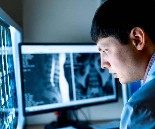
"Radiologists are uniquely positioned to educate other healthcare providers on how to properly remove personal health information from radiologic imaging files," wrote Stern and colleague William Weadock, MD, also of the university. PowerPoint presentations that include medicalimages are a valuable educational tool, Stern and Weadock noted.
Additionally, the staff should include certified radiologic technologists and board-certified radiologists who specialize in interpreting various imagingmodalities. Ensuring your imaging center has these certifications guarantees that your scans will be handled by knowledgeable and experienced professionals.
Teleradiology & Radiology data for artificial intelligence (AI) Introduction: Embark on a journey into the world of medicalimaging as we unravel the distinctions between two powerful diagnostic tools—Computed Tomography (CT) scans and Positron Emission Tomography (PET) scans.
Medicalimaging has evolved over centuries, starting with X-rays in 1895, progressing to CT, MRI, and PETscans. The post Revolutionising Healthcare: A Historical Perspective on MedicalImaging appeared first on Open Medscience.
Hospitals and medical practices of all sizes, no matter their target demographic or geographical location, often struggle to ensure their patients have access to all of the essential imagingmodalities necessary to diagnose a wide variety of medical conditions.
PETscans are crucial for detecting metabolic activity, providing valuable insights into cancer, neurological disorders, and cardiovascular diseases. The post How to Read a PETScan: A Basic Understanding appeared first on Open MedScience.
Medicalimaging crucially enhances oncology, aiding early cancer detection and effective treatment planning. The post The Transformative Impact of MedicalImaging in Oncology appeared first on Open Medscience.
This description highlights the significant advancements in medicalimaging that are transforming healthcare. Advanced algorithms can analyze images more quickly and accurately, aiding in early diagnosis and treatment planning. This aids in the early detection and monitoring of diseases.
Advances in medicalimaging technology have significantly improved diagnostic accuracy, enabling earlier detection and more personalised treatments. The post Advances in MedicalImaging Technology: Unlocking the Mysteries of the Human Body appeared first on Open MedScience.
PACS – Picture Archiving and Communication System; a system involved in acquiring the medicalimages, transmission, viewing, storage, and retrieval of same images. of imagingmodalities, storage space, no. Basically, PACS is an electronic version of the file room and reading room for radiologists.
DICOM Image Format is an international standard to transmit, store, retrieve, print, process, and display medicalimaging information. DICOM allows transmitting medicalimaging data to devices like scanners, servers, workstations, printers, network hardware, and PACS.
Positron Emission Tomography Imaging has advanced with cutting-edge technologies, enhancing diagnostic accuracy and expanding clinical applications dramatically. The post Breakthroughs in Positron Emission Tomography Imaging appeared first on Open MedScience.
We organize all of the trending information in your field so you don't have to. Join 5,000 users and stay up to date on the latest articles your peers are reading.
You know about us, now we want to get to know you!
Let's personalize your content
Let's get even more personalized
We recognize your account from another site in our network, please click 'Send Email' below to continue with verifying your account and setting a password.


Let's personalize your content