Remove Imaging Modalities Remove Picture Archiving and Communication System Remove Ultrasound
article thumbnail

PACS: Diminishing the Distance and Enabling the Real-Time Services in Teleradiology

Cloudex Radiology

PACS – Picture Archiving and Communication System; a system involved in acquiring the medical images, transmission, viewing, storage, and retrieval of same images. The fundamental parts of PACS are – imaging acquisition, display workstations, archive servers.

PACS 52
article thumbnail

Teleradiology Integration: Harmonizing Imaging Technologies for Better Healthcare (Chhattisgarh, Kabirdham, Gariaband, Surajpur, Bijapur, Bemetara, Kanker, Sarangarh-Bilaigarh, Bilaspur)

Future Teleradiology

This blog post explores how the seamless incorporation of teleradiology into healthcare systems leads to improved diagnostics, streamlined workflows, and ultimately, enhanced patient care. Interconnected Imaging Modalities: Discuss the integration of diverse imaging modalities in teleradiology.

Insiders

Sign Up for our Newsletter

This site is protected by reCAPTCHA and the Google Privacy Policy and Terms of Service apply.

Trending Sources

article thumbnail

What is CPACS in Cardiology?

Intelerad

CPACS stands for Cardiovascular Picture Archive and Communication System. It is a medical imaging system used to store, manage, and share cardiology images. Cardiologists use CPACS systems to view, diagnose, and treat heart conditions. How Does CPACS Work?

article thumbnail

A brief history of radiology

Radiology Cafe

The theoretical basis for ultrasound physics has been around since 1794, but it wasn’t until 1942, when Dr Karl Theodore Dussik in Austria transmitted an ultrasound beam through a human skull to view the brain, that ultrasound was first used in medicine. (13) This was a defining publication in the field of medical ultrasound. (14)

article thumbnail

DEVELOPMENT OF HEPATOCELLULAR CARCINOMA

Cloudex Radiology

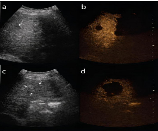
DIAGNOSIS OF HEPATOCELLULAR CARCINOMA IMAGING MODALITIES ULTRASOUND: Small focal HCC appears hypoechoic compared with normal liver. All image data were transmitted directly to our picture archiving and communication system. Sagittal and coronal reformats of images were also obtained(10).