This site uses cookies to improve your experience. To help us insure we adhere to various privacy regulations, please select your country/region of residence. If you do not select a country, we will assume you are from the United States. Select your Cookie Settings or view our Privacy Policy and Terms of Use.
Cookie Settings
Cookies and similar technologies are used on this website for proper function of the website, for tracking performance analytics and for marketing purposes. We and some of our third-party providers may use cookie data for various purposes. Please review the cookie settings below and choose your preference.
Used for the proper function of the website
Used for monitoring website traffic and interactions
Cookie Settings
Cookies and similar technologies are used on this website for proper function of the website, for tracking performance analytics and for marketing purposes. We and some of our third-party providers may use cookie data for various purposes. Please review the cookie settings below and choose your preference.
Strictly Necessary: Used for the proper function of the website
Performance/Analytics: Used for monitoring website traffic and interactions
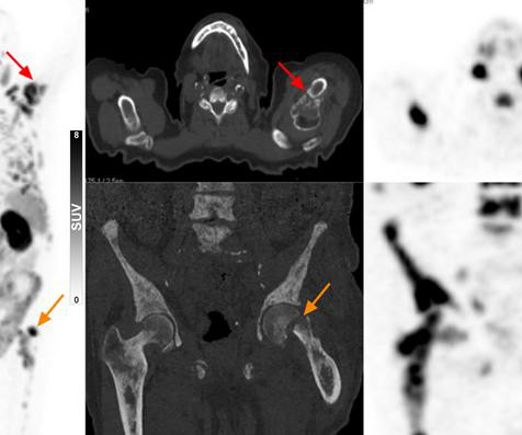
Although the most common site of MBC is bone, there is currently no standardized imagingmodality that offers accurate assessment of bone treatment response, Masperi and Girlando explained. Data on radiation doses and the administration of contrast agents or radiopharmaceuticals were also collected.
A team of researchers at Boston Children’s Hospital has developed an age-specific dose catalog for estimating radiation exposure to children from diagnostic and interventional radiology fluoroscopy procedures.
Currently, CT and MRI are the imagingmodalities of choice for assessing diagnosed patients, yet have been shown to miss small lesions or metastatic deposits, the authors explained. Hepatocellular carcinoma (HCC) is the third most common cause of cancer-related deaths worldwide, accounting for over 800,000 deaths annually.
Contrast-enhanced mammography (CEM) has a slightly higher mean glandular dose (MGD) than other breast imagingmodalities, according to research published January 15 in the American Journal of Roentgenology. Mean glandular dose (MGD) of breast imagingmodalities Mammography DBT Combined mammography-DBT CEM (overall) Median MGD (mGy) 4.07
Screening with breast MRI is "recommended for very high-risk patients, such as BRCA mutation carriers, as they begin screening at a young age and may be more susceptible to the negative effects of ionizing radiation," the authors wrote. Breast MRI is effective for imaging patients with implants, as it can assess the integrity of the implants.
They found that F-18 FDG-PET/CT can be useful in breast cancer management, including initial staging, assessing neoadjuvant systemic treatment response, assessing treatment response in the metastatic setting, searching for loco-regional or metastatic recurrence, and re-staging after therapy, as well as radiation therapy planning.
While medical imaging has proven its value in health screening, such procedures can be anxiety-inducing and uncomfortable for patients. This includes patients experiencing claustrophobia in MRI machines, compression pain from mammography, or concerns about radiation exposure.
Prostate artery embolization (PAE) is a growing minimally invasive treatment for benign prostatic hyperplasia (BPH), with imagingmodality selection playing a critical role in determining procedural radiation exposure.
Food and Drug Administration (FDA) has said CT is still the preferred imagingmodality for patients with medical devices. In an October 15 communication , the agency said it had received reports of electronic medical devices being damaged during CT scans due to radiation.
milla1cf Tue, 06/13/2023 - 20:02 June 13, 2023 — The Cancer Institute at Northwell Lenox Hill Hospital is the first in New York State to use the HyperSight imaging solution, which employs state-of-the-art cone-beam computed tomography technology to deliver faster, sharper images during the radiation therapy treatment process.
PEM is one molecular breast imaging method that has shown promise in lowering the number of false-positive cases, owing to its superior specificity over MRI. However, PEM’s higher radiation dose has steered radiologists away from using the modality.
Even if some private urology, radiation oncology practices, or radiologist groups are building the ability to perform theranostics, experts are cautious about patient management, radiation safety, and the risk of unnecessary imaging. However, few freestanding theranostics centers exist today.
Geoffrey Rubin, MD, of the University of Arizona College of Medicine Tucson, offered session attendees an overview of key issues in the radiology workforce landscape, highlighting a 2024 consensus committee report from the American Society of Radiologic Technologists (ASRT) on the future of medical imaging and radiation therapy.
Ultrasound proponents will have the opportunity to present and see the modality’s versatility in helping detect and diagnose pathologies such as liver cancer, breast cancer, and thyroid cancer. They can also highlight the resulting benefits that are possible, of course, without the ionizing radiation of some other imagingmodalities.
All in all, ultrasound is a versatile modality that can help detect and diagnose pathologies such as liver cancer, breast cancer, and thyroid cancer, with RSNA presentations aiming to showcase these benefits that are possible, of course, without the ionizing radiation of some other imagingmodalities.
The need for imagingmodalities to support an earlier, more accurate diagnosis continues to drive AI applications in the medical imaging market. In fact, the size of the market for AI in medical imaging is experiencing phenomenal growth, expected to increase from $1.12 billion in 2022 to $27.52
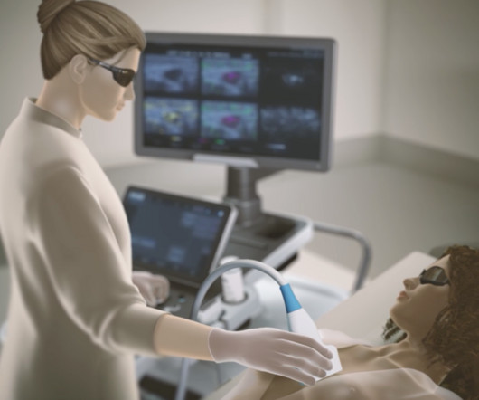
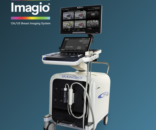
The result is a diagnostic imagingmodality that delivers functional information regarding suspicious breast masses, increasing confidence regarding the need for invasive breast cancer diagnostic biopsies.
Today, Low Dose Computed Tomography (LDCT) imaging is the gold standard for detecting abnormalities in the lungs because it exposes patients to less radiation than a traditional CT scan. Since LDCTs take images from several different angles, they provide a 3D picture of a selected structure for precision diagnostics.
mtaschetta-millane Tue, 07/02/2024 - 09:53 July 2, 2024 — A new editorial paper was published in Oncoscience ( Volume 11 ) on May 20, 2024, entitled, “ Deep learning-assisted lesion segmentation in PET/CT imaging: A feasibility study for salvage radiation therapy in prostate cancer.” In this new editorial, researchers Richard L.J.
"The inequity in access to detailed stroke imaging highlights a potential role for dedicated ultralow-field-strength ( Patients presenting with stroke need rapid diagnosis, and researchers have continued to develop imagingmodalities that don't impart high levels of radiation and, in the case of MRI, have low magnetic field strength.
Key Points: The most important advantage of weight bearing CT (WBCT), which utilizes cone beam CT (CBCT) technology (a three-dimensional (3D) imagingmodality) is immediate access to 3D images, resulting in faster and better diagnostic capabilities.
MRI-Scan-Teleradiology Introduction: The advancement of medical imaging technologies has revolutionized healthcare diagnostics, but concerns about radiation exposure persist. Optimizing Imaging Protocols: Tailoring for Precision: Discuss the significance of optimizing imaging protocols for each patient.
X-ray and CT imaging are the go-to exams for trauma, Fritz conceded, but MRI is gaining ground as an effective modality for MSK trauma applications, in part because it does not expose patients to radiation and because it can image soft tissue well. All images and captions courtesy of Prof. Benjamin Fritz, MD.
Since almost half of the screening population has dense breasts, many of these patients require additional breast imaging, often with MRI , after mammography. Low-dose positron emission mammography (PEM) is a novel molecular imaging technique that provides improved diagnostic performance at a radiation dose comparable to that of mammography.
Understanding the distinction between radiography and sonography is vital in the increasingly specialized and multifaceted world of medical imaging. Radiography uses ionizing radiation to capture detailed internal images, which helps diagnose various conditions.
Through the appearance or absence of two hallmark indicators of cancer — angiogenesis and deoxygenation — the Imagio OA/US Breast Imaging System is a more effective tool to help radiologists confirm or rule out malignancy compared with traditional diagnostic imagingmodalities.
At the same time, the use of imaging, particularly CT, has been on the rise in emergency departments and may present health risks due to the effects of radiation exposure. As a result, the researchers sought to assess whether HIE adoption could lead to a decline in repeat imaging in EDs.
Section 1: The Shifting Paradigm in Trauma Imaging: Introduce the changing dynamics in trauma radiology, highlighting the transition from traditional imaging approaches to the emergence of extraneous imagingmodalities.
Published in the July issue of The Journal of Nuclear Medicine , this new research demonstrates how imaging with PSMA PET/CT can potentially reduce the number of prostate biopsies and associated complications in the elderly while providing accurate staging data.
The balance of dose and image quality is even more important in pediatric medical imaging. Not only are children more radiosensitive than adults (the cancer risk per unit dose of ionizing radiation is higher), but children also have a longer expected lifetime, which puts them at greater risk of cancer following radiation exposure.(1)
Continuous-rotation computed tomography (CT) fluoroscopy is an imagingmodality widely used in interventional radiology (IR) procedures, facilitating precise punctures even into small lesions and lesions deep within the body by rapid, real-time, and high-resolution tomographic images (1,2).
Teleradiology & Radiology data for artificial intelligence (AI) Introduction: Embark on a journey into the world of medical imaging as we unravel the distinctions between two powerful diagnostic tools—Computed Tomography (CT) scans and Positron Emission Tomography (PET) scans.
AI can be leveraged to make incremental improvements at every stage of the medical imaging pipeline, beyond the tasks well-suited for a large language model. Transparency measures include disclosing study design, sample size, demographic characteristics, equipment specifications, imagingmodality, target population, and target use case.
Advanced ImagingModalities : Emerging imagingmodalities, including MRI, CT, PET/CT, and ultrasound, offer a comprehensive view of the human body, aiding in the detection and management of complex medical conditions.
Highlight its pivotal role in diagnosing medical conditions by capturing internal images of the body. Ionizing Radiation: Understanding the Nature of X-Rays: Explain the concept of ionizing radiation and how X-rays fit into this category. Discuss the potential biological effects of ionizing radiation on tissues.
Discuss how this approach delivers radiation with exceptional precision to cancer cells. Advances in Cardiac Imaging: Precision in Heart Disease Diagnosis: Highlight recent breakthroughs in cardiac imaging within nuclear medicine. Discuss how these combinations provide comprehensive anatomical and functional information.
Advanced algorithms can analyze images more quickly and accurately, aiding in early diagnosis and treatment planning. 3D and 4D Imaging : The development of 3D and 4D imaging techniques provides a more comprehensive and dynamic view of internal structures, allowing for improved anatomical understanding and better treatment planning.
Studies show that neuromelanin MRI may provide useful adjunctive information to aid in the evaluation of Parkinson’s disease, and avoids the disadvantages of current adjunctive imagingmodalities such as DaT-SPECT, which requires a five-hour procedure, intravenous radiation exposure, and can cost well over $3,000 [1-6].
In a recent paper published in Foot and Ankle Clinics, authors Alessio Bernasconi and Francois Lintz even suggest weight bearing CT should be the first-line imagingmodality for cavovarus patients because of the advantages it offers for surgical planning.
Today, the same should be said of AI ROI image-guided technology and the radiation reduction and protection it provides. Instead of a constant beam of X-ray energy, pulsed fluoro technology provided an innovation that reduced radiation exposure by ~65% from non-pulsed systems. The same with flat panels.
Introduction to Radiology : Radiology is a branch of medicine that uses medical imaging techniques to diagnose and treat diseases and injuries. It includes various imagingmodalities such as X-rays, CT scans, MRIs, ultrasounds, and nuclear medicine.
Digital Transformation : Traditional film-based radiography is being replaced by digital imaging systems, which offer higher resolution, more efficient data management, and the ability to share images electronically.
MRI Imaging of Prostate was started sometime during the mid-1980’s. In mpMRI, the conventional T1w and T2w imaging is clubbed with atleast one of the functional MR imaging techniques like Diffusion Weighted Imaging (DWI), Dynamic Contrast Enhanced -MRI, MR-Spectroscopy etc….
We organize all of the trending information in your field so you don't have to. Join 5,000 users and stay up to date on the latest articles your peers are reading.
You know about us, now we want to get to know you!
Let's personalize your content
Let's get even more personalized
We recognize your account from another site in our network, please click 'Send Email' below to continue with verifying your account and setting a password.


Let's personalize your content