This site uses cookies to improve your experience. To help us insure we adhere to various privacy regulations, please select your country/region of residence. If you do not select a country, we will assume you are from the United States. Select your Cookie Settings or view our Privacy Policy and Terms of Use.
Cookie Settings
Cookies and similar technologies are used on this website for proper function of the website, for tracking performance analytics and for marketing purposes. We and some of our third-party providers may use cookie data for various purposes. Please review the cookie settings below and choose your preference.
Used for the proper function of the website
Used for monitoring website traffic and interactions
Cookie Settings
Cookies and similar technologies are used on this website for proper function of the website, for tracking performance analytics and for marketing purposes. We and some of our third-party providers may use cookie data for various purposes. Please review the cookie settings below and choose your preference.
Strictly Necessary: Used for the proper function of the website
Performance/Analytics: Used for monitoring website traffic and interactions
In the third blog of her series on AI and the radiographer, Shamie Kumar explores the impact on the radiographer when AI is integrated within an imagingmodality. The question to explore in this blog is when AI is integrated within an imagingmodality itself and how that may impact a radiographer.
Kim Mason Kim Mason, an Audit and Research Radiographer for Mid Yorkshire Teaching Hospitals Trust, talks about their role as well as the value of radiographer engagement in research activities and how to get involved. Hi, I’m Kim and I am an alternative-styled, funky-haired, septum-pierced, disabled Audit and Research Radiographer.
The need for imagingmodalities to support an earlier, more accurate diagnosis continues to drive AI applications in the medical imaging market. In fact, the size of the market for AI in medical imaging is experiencing phenomenal growth, expected to increase from $1.12 billion in 2022 to $27.52
The enduring shortage is affecting staffing for radiographers and radiologists; and all imagingmodalities. Marion Anderson, CRA, Diagnostic Imaging Manager at Karmanos Cancer Center in Detroit, Michigan reports that her facility is down two-thirds of staff for CT and mammography exams. Bureau of Labor Statistics.
Advanced algorithms can analyze images more quickly and accurately, aiding in early diagnosis and treatment planning. 3D and 4D Imaging : The development of 3D and 4D imaging techniques provides a more comprehensive and dynamic view of internal structures, allowing for improved anatomical understanding and better treatment planning.
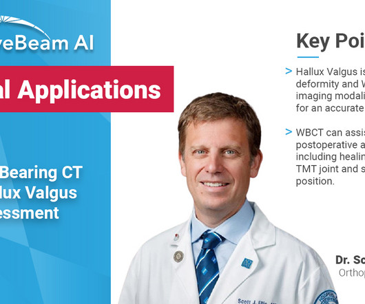
Key Points: Weight bearing CT (WBCT) can detect signs of osteoarthritis (OA), such as osteophytes, subchondral cysts, and joint space narrowing better than radiographs. Emerging evidence suggests WBCT reveals meniscal extrusion not detected by MRI and WBCT arthrography visualizes meniscal tears.
Diagnosis While radiographs are typically sufficient to make the diagnosis, WBCT scans may be useful to plan surgical treatment. Accurately assess sesamoid position as plain radiographs cannot determine whether the sesamoids have been reduced within their grooves 5. Accurately assess healing in the 1st TMT joint 4. 10.30795/jfootankle.2022.v16.1674.
Reading Time: 4 minutes read Key takeaways from the 2023 RSNA Conference The topic of artificial intelligence technology trends in medical imaging was once again infused throughout sessions and on display in the exhibitor hall at the 2023 Radiological Society of North America (RSNA) scientific assembly and annual meeting.
Likewise, it is in the best interest of the child to decline an order for an exam for scoliosis if you only have conventional radiography equipment and are incapable of offering the child the benefit of decreased exposure by standard PA positioning and digital imaging systems.
Trauma : In these injuries one would normally go for a standard radiography followed by second-line imaging such as CT. Trauma : WBCT imaging is my preferred imagingmodality as well for ankle sprain, foot sprain, as well as any possible associated fractures.
We organize all of the trending information in your field so you don't have to. Join 5,000 users and stay up to date on the latest articles your peers are reading.
You know about us, now we want to get to know you!
Let's personalize your content
Let's get even more personalized
We recognize your account from another site in our network, please click 'Send Email' below to continue with verifying your account and setting a password.
Let's personalize your content