This site uses cookies to improve your experience. To help us insure we adhere to various privacy regulations, please select your country/region of residence. If you do not select a country, we will assume you are from the United States. Select your Cookie Settings or view our Privacy Policy and Terms of Use.
Cookie Settings
Cookies and similar technologies are used on this website for proper function of the website, for tracking performance analytics and for marketing purposes. We and some of our third-party providers may use cookie data for various purposes. Please review the cookie settings below and choose your preference.
Used for the proper function of the website
Used for monitoring website traffic and interactions
Cookie Settings
Cookies and similar technologies are used on this website for proper function of the website, for tracking performance analytics and for marketing purposes. We and some of our third-party providers may use cookie data for various purposes. Please review the cookie settings below and choose your preference.
Strictly Necessary: Used for the proper function of the website
Performance/Analytics: Used for monitoring website traffic and interactions
A team led by PhD candidate Lu Zhang of Shanghai Jiao Tong University in Shanghai, China, developed a large language model (LLM) that generates interpretations (“impressions”) on reports based on imaging findings and evaluated its performance in professional and linguistic dimensions.
In the third blog of her series on AI and the radiographer, Shamie Kumar explores the impact on the radiographer when AI is integrated within an imagingmodality. The question to explore in this blog is when AI is integrated within an imagingmodality itself and how that may impact a radiographer.
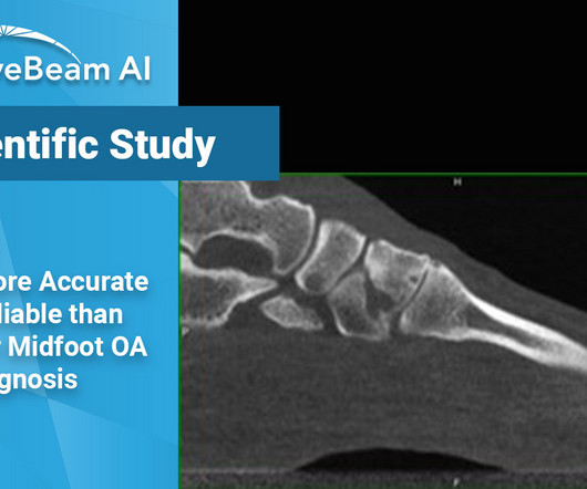
Key Points: When evaluating midfoot arthritis osteoarthritis (OA), Weight Bearing X-Ray shows many false negatives and false positives, even when read by a musculoskeletal (MSK) radiologist, as compared to weight bearing CT (WBCT). Researchers used internal data from a cohort of 302 patient feet.
Our portable X-ray equipment provides high quality diagnostic imaging capabilities for evaluating injury and illness. “This addition helps round out our care offerings,” says Jeanne Walter, vice president of marketing and sales at MinXray.
Introduction: The history of X-rayimaging is a testament to the unceasing march of technology in healthcare. From the days of photographic film to the digital age, this blog traces the remarkable evolution of X-rayimaging, shedding light on how technology has transformed the practice of medicine.
Teleradiology Introduction: X-ray technology has been a cornerstone of modern medicine for over a century. This blog explores the evolution, significance, and the latest advancements in X-ray technology, shedding light on how it continues to shape and revolutionize the healthcare industry.
The need for imagingmodalities to support an earlier, more accurate diagnosis continues to drive AI applications in the medical imaging market. In fact, the size of the market for AI in medical imaging is experiencing phenomenal growth, expected to increase from $1.12 billion in 2022 to $27.52
In December, 1895, Wilhelm Röntgen would x-ray the hand of Anna Bertha Ludwig, his wife, using a photographic plate. The new discovery lit a fire in the scientific community, and was so sensational that in the following year over 1,000 articles would be published on the topic of X-rays. That is where I come in.
Konica Minolta Healthcare Americas extends this history of innovation with a keen focus on contributing to life-changing advances through the transformation of primary imaging, including digital radiography (DR), ultrasound and imaging IT solutions. "At
Highlights of DIS’s presence at VMX 2024 include: Advanced Imaging Solutions: DIS will showcase a range of advanced diagnostic imaging systems designed to meet the diverse needs of veterinary practices. The post Cutting-Edge Veterinary Imaging Solutions at VMX Veterinary Meeting & Expo appeared first on Vet X-ray.
Closeup of X-ray photography of human brain Description: The field of radiology is in the midst of a remarkable revolution, driven by cutting-edge technologies and innovations. This description highlights the significant advancements in medical imaging that are transforming healthcare.
Technology in Radiology : Radiology relies on a sophisticated array of imaging technologies, such as X-rays, CT scans, MRIs, and ultrasound, to reveal the complexities of human anatomy and pathology.
Likewise, it is in the best interest of the child to decline an order for an exam for scoliosis if you only have conventional radiography equipment and are incapable of offering the child the benefit of decreased exposure by standard PA positioning and digital imaging systems. Reference: 1 1 U.S.
Historically, the chest x-ray (CXR) was the initial imagingmodality used during the primary survey to look for pneumothorax (PTX) but it is notoriously insensitive. Patients without confirmatory CT chest imaging were excluded which creates a massive selection bias. Acad Emerg Med. 2005 Sep;12(9):844-9.
We organize all of the trending information in your field so you don't have to. Join 5,000 users and stay up to date on the latest articles your peers are reading.
You know about us, now we want to get to know you!
Let's personalize your content
Let's get even more personalized
We recognize your account from another site in our network, please click 'Send Email' below to continue with verifying your account and setting a password.


Let's personalize your content