This site uses cookies to improve your experience. To help us insure we adhere to various privacy regulations, please select your country/region of residence. If you do not select a country, we will assume you are from the United States. Select your Cookie Settings or view our Privacy Policy and Terms of Use.
Cookie Settings
Cookies and similar technologies are used on this website for proper function of the website, for tracking performance analytics and for marketing purposes. We and some of our third-party providers may use cookie data for various purposes. Please review the cookie settings below and choose your preference.
Used for the proper function of the website
Used for monitoring website traffic and interactions
Cookie Settings
Cookies and similar technologies are used on this website for proper function of the website, for tracking performance analytics and for marketing purposes. We and some of our third-party providers may use cookie data for various purposes. Please review the cookie settings below and choose your preference.
Strictly Necessary: Used for the proper function of the website
Performance/Analytics: Used for monitoring website traffic and interactions
"Nonradiologist specialties, aside from cardiology, lack the rigorous and comprehensive training in imaging interpretation that occurs during the four years of a radiology residency program. million office-based imaging claims for Medicare fee-for-service beneficiaries in 2022 that were ordered by nonradiologists.
Higher Medicaid-to-Medicare reimbursement ratios (MMRR) are linked to increased likelihood of Medicaid patients receiving CT, MR, ultrasound, and x-rayimaging, researchers have reported. Ultrasound 0.85 X-ray or fluoroscopy 0.82 higher for ultrasound, and 31.8% higher for x-ray.
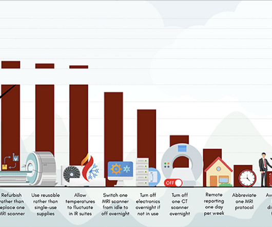
The researchers also suggested that decision-support tools can be implemented to choose lower-energy imaging tests when appropriate. Carbon dioxide emissions vary by imagingmodality and are higher for MRI and CT compared with ultrasound and x-rays.
They evaluated imaging referral completion rates in their urban free clinic for underserved patients, including associated factors. Of the modalities included in the study, ultrasound had a completion rate of 59%, followed by 45% for x-ray and 37% for mammography.
The work included data from eight Google searches related to a medical imagingmodality such as x-rays, CT scans, MRIs, PET scans, CT and MR angiography, and ultrasounds. A search for "Radiology Chest X-ray filetype:ppt" resulted in accessible PHI in 40% of the presentations with images.
Substituting less energy-consuming ultrasound for x-ray or CT reduced energy use by as much as 8% during diagnostic radiology processes and 31.2% during indirect radiology department activities, according to findings of a pilot study presented March 1 at ECR 2024. kilowatts per patient, Masperi said.
Furthermore, image fidelity between original and rendered images has traditionally been evaluated using pixel-based metrics like MSE and PSNR, which prioritize mathematical precision and often fall short of capturing perceptual or diagnostic relevance. However, more than three-quarters of AI-based medical devices authorized by the U.S.
Adapters can be utilized to streamline workflows in radiology and other image-generating specialties, enabling, for example, researchers to build models to automatically route imaging scans to specialists, or flag potential abnormalities for further review, Microsoft said.
Adopting health information exchange (HIE) between unaffiliated healthcare providers leads to significant decreases in repeat imaging studies in emergency departments (EDs) and could potentially save millions of dollars, according to a large study from the University of Michigan. of ultrasound cases), and 29,073 repeat chest x-rays (19.5%
In 2024, the top three diagnostic imagingmodalities used for cardiology-related imaging exams are SPECT, CT, and ultrasound, according to IMV's recently published cross-modality report 2024 Cardiology in Diagnostic Imaging Market Outlook Report. Davin Korstjens of IMV Medical Information Division.
a key global supplier of portable, compact digital imaging equipment, has added a line of wireless handheld ultrasound technology to speed diagnosis and improve triage efficiency in medical care. Our portable X-ray equipment provides high quality diagnostic imaging capabilities for evaluating injury and illness. “Our
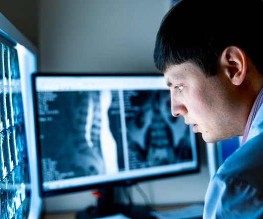
Radiologists are medical doctors who specialize in interpreting imaging studies like X-rays, CT scans, MRIs, and ultrasounds to diagnose and guide treatment for various conditions. This rigorous training covers all imagingmodalities, from X-rays to advanced techniques like MRI and PET/CT scans.
Using a dedicated MRI system in the ED offers many potential benefits, from improving access to the modality for trauma patients to offering a more certain diagnosis than other imagingmodalities can, said presenter Catherine Mandel, MD, of the University of Melbourne in Australia. The bottom line?
Hospitals and medical practices of all sizes, no matter their target demographic or geographical location, often struggle to ensure their patients have access to all of the essential imagingmodalities necessary to diagnose a wide variety of medical conditions.
Additionally, the staff should include certified radiologic technologists and board-certified radiologists who specialize in interpreting various imagingmodalities. Ensuring your imaging center has these certifications guarantees that your scans will be handled by knowledgeable and experienced professionals.
Carolina Breast Imaging Specialists, PLLC in Greenville, North Carolina intends to change that as the first radiology center in the U.S.A. to adopt the recently available Imagio Breast Imaging System. And it does this without exposing patients to potentially harmful ionizing radiation ( x-rays ) or contrast agents.
Bracco Imaging S.p.A., part of the Bracco Group, is a global diagnostic imaging provider, headquartered in Milan, Italy, which develops, manufactures and markets diagnostic imaging agents and solutions.
Foundation models for medical imaging could help, but will be hard to create. Radiologists can identify hundreds of different types of diseases across many imagemodalities (MRI, CT, chest X-ray, other X-ray, mammography, ultrasound, PET, SPECT) and also have a detailed knowledge of what variations of anatomy are normal vs anomalous.
Medical imaging is a crucial tool in modern healthcare, providing detailed visuals of the human body’s internal structures and helping in the accurate diagnosis and treatment of various conditions. Magnetic Resonance Imaging (MRI) MRI stands for Magnetic Resonance Imaging. X-rays are fast, painless, and commonly used.
Which of the following imagingmodalities is indicated at this time? Barium contrast X-ray CT scan Magnetic resonance imagingUltrasound FOR THE RIGHT ANSWER CLICK ON THE ROSH REVIEW LOGO BELOW References Burkart JM, Bleyer A. McGraw Hill; 2020:(Ch) 90. link] Szeto CC, Li PKT. Clin J Am Soc Nephrol.
Another potential action is to implement decision-support tools to choose lower-energy imaging tests when appropriate. Carbon dioxide equivalent, or CO2e emissions, vary by imagingmodality and are higher for MRI and CT compared to ultrasound and X-rays.
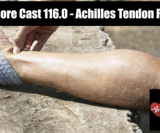
Partial rupture of the proximal Achilles tendon: a differential diagnostic problem in ultrasoundimaging. Rezaie, MD (Twitter/X: @srrezaie ) The post REBEL Core Cast 116.0 – Achilles Tendon Rupture appeared first on REBEL EM - Emergency Medicine Blog. Total Achilles tendon rupture. Sports Med. 1998; 25(2): 79-100.
Konica Minolta Healthcare Americas extends this history of innovation with a keen focus on contributing to life-changing advances through the transformation of primary imaging, including digital radiography (DR), ultrasound and imaging IT solutions. "At
Background: The use of ultrasound is well established for trauma patients in the emergency department, with almost every patient receiving a FAST (Focused Assessment with Sonography in Trauma) examination as part of the “ABC’s” of trauma. Not so FAST- Chest ultrasound underdiagnoses traumatic pneumothorax. PMID: 34932040.
We recently adopted a nationwide pediatric appendix ultrasound performance protocol, sonographer worksheet, and radiologist reporting template in order to decrease CT utilization for this diagnosis nationwide. You can enroll in the free, on-demand course at Pediatric Appendix Ultrasound Standardized Performance and Reporting Training.
Highlights of DIS’s presence at VMX 2024 include: Advanced Imaging Solutions: DIS will showcase a range of advanced diagnostic imaging systems designed to meet the diverse needs of veterinary practices. The post Cutting-Edge Veterinary Imaging Solutions at VMX Veterinary Meeting & Expo appeared first on Vet X-ray.
PACS works as a host that integrates the radiological images acquired from different radiological imagingmodalities (X-Ray, Ultrasound, CT, MRI , PET Scan, Nuclear Medicine, etc…) with a network of information system (RIS and or HIS), EMR, different work stations and image storage/archival system.
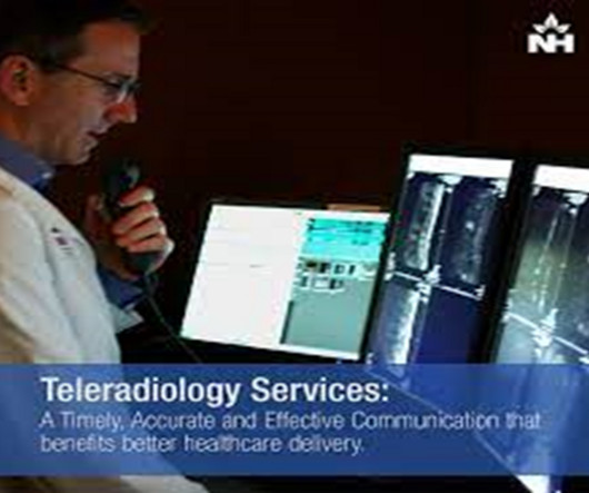
Interconnected ImagingModalities: Discuss the integration of diverse imagingmodalities in teleradiology. Explore how systems are designed to handle data from modalities such as X-ray, CT, MRI, and ultrasound, providing a comprehensive diagnostic overview.
The Whimsy of Modalities: Magic in Every Image Explore the whimsical nature of various imagingmodalities, each possessing its own magical properties within the mad multiverse. Discuss how the peculiar doctor harnesses the magic of MRI, CT, X-ray, and ultrasound to unveil hidden realms.
Technology in Radiology : Radiology relies on a sophisticated array of imaging technologies, such as X-rays, CT scans, MRIs, and ultrasound, to reveal the complexities of human anatomy and pathology.
A normal lactate does not rule out the diagnosis Plain X-rays perform poorly in making or ruling out the diagnosis. 2.3 – 5.4) (-) LR: 0.18 (0.09 – 0.35) Ultrasound Findings Dilated loops of bowel (diameter > 2.5 Abdominal X-ray 75% 66% 1.6 Absent bowel sounds Peritoneal signs (i.e. 0.27 – 0.83
Introduction to Radiology : Radiology is a branch of medicine that uses medical imaging techniques to diagnose and treat diseases and injuries. It includes various imagingmodalities such as X-rays, CT scans, MRIs, ultrasounds, and nuclear medicine.
In December, 1895, Wilhelm Röntgen would x-ray the hand of Anna Bertha Ludwig, his wife, using a photographic plate. The new discovery lit a fire in the scientific community, and was so sensational that in the following year over 1,000 articles would be published on the topic of X-rays. That is where I come in.
Different expertise is needed to interpret images generated from different technologies, such as X-ray, MRI, CT, PET, ultrasound, mammography, and DEXA. Schedule a meeting with Real Radiology Benefit From Our Wide-Ranging Expertise The scope of radiology varies widely.
Introduction of X-ray pneumoencephalography in 1947 vastly improved surgeons’ ability to localize targets, particularly with the later development of detailed stereotactic atlases. The CT and MRI images are post-contrast images and detailed planning is done to use a trajectory, which does not go through the ventricles, blood vessels.
This standard has revolutionized the radiology industry, encompassing many imagingmodalities such as X-rays, computed tomography (CT), magnetic resonance imaging (MRI), ultrasound, nuclear medicine, PET scans, etc.
To excel in medical imaging, one must pursue postgraduate education beyond basic nursing or medicine. The post A Guide To Education and Upskilling For Professionals in The Medical Imaging Field appeared first on Open Medscience.
It all started when Wilhelm Conrad Röntgen discovered X-rays in 1895. After working for weeks in his lab experimenting on the production of ‘strange rays’, which he referred to as ‘X’, he asked his wife Anna Bertha to lend ‘a hand’, the left one to be precise, which he used to produce the first X-rayimage.
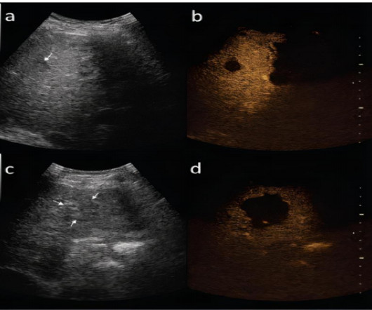
DIAGNOSIS OF HEPATOCELLULAR CARCINOMA IMAGINGMODALITIESULTRASOUND: Small focal HCC appears hypoechoic compared with normal liver. Technical parameters were X-ray tube current 160 to 220 mA; tube voltage 120 kV; collimation 5mm; rotation speed 0.75 Tumour thrombus may be visible.(8) s; matrix 512×512.
Newborns' livers can be affected by a variety of congenital and acquired diseases, and imaging plays an important role in the workup and management of these, according to a study published November 7 in RadioGraphics. The team outlined the pros and cons of various modalities for neonatal liver imaging: X-ray.
We organize all of the trending information in your field so you don't have to. Join 5,000 users and stay up to date on the latest articles your peers are reading.
You know about us, now we want to get to know you!
Let's personalize your content
Let's get even more personalized
We recognize your account from another site in our network, please click 'Send Email' below to continue with verifying your account and setting a password.


Let's personalize your content