This site uses cookies to improve your experience. To help us insure we adhere to various privacy regulations, please select your country/region of residence. If you do not select a country, we will assume you are from the United States. Select your Cookie Settings or view our Privacy Policy and Terms of Use.
Cookie Settings
Cookies and similar technologies are used on this website for proper function of the website, for tracking performance analytics and for marketing purposes. We and some of our third-party providers may use cookie data for various purposes. Please review the cookie settings below and choose your preference.
Used for the proper function of the website
Used for monitoring website traffic and interactions
Cookie Settings
Cookies and similar technologies are used on this website for proper function of the website, for tracking performance analytics and for marketing purposes. We and some of our third-party providers may use cookie data for various purposes. Please review the cookie settings below and choose your preference.
Strictly Necessary: Used for the proper function of the website
Performance/Analytics: Used for monitoring website traffic and interactions
A team led by Junqi Han, MD, from the Affiliated Hospital of Qingdao University in China found that its model combining data from mammography images, ultrasound images, and other characteristics performed well in predicting disease-free survival of breast cancer. This means the combined model could predict prognosis after surgery.
VIENNA -- It's definitely possible to develop a successful cardiac imaging practice, according to a professional development presentation delivered February 28 at the ECR in Vienna. Gutberlet described how he came to cardiac imaging early in his medical career. years of follow-up compared with CT imaging (2.1% But he persevered.
The need for imagingmodalities to support an earlier, more accurate diagnosis continues to drive AI applications in the medical imaging market. In fact, the size of the market for AI in medical imaging is experiencing phenomenal growth, expected to increase from $1.12 billion in 2022 to $27.52
In the third blog of her series on AI and the radiographer, Shamie Kumar explores the impact on the radiographer when AI is integrated within an imagingmodality. The question to explore in this blog is when AI is integrated within an imagingmodality itself and how that may impact a radiographer.
The green project conducted by radiographers at the European Institute of Oncology in Milan and nearby Hospital of Legnano saved an estimated 12,000 euros ($13,000), said Andrea Masperi, who presented the details. The group developed a "green imaging review" for patients accessing the emergency room.
Mitigating radiology workforce strain requires revisioning the specialty and adding new roles to manage imaging demand, according to a presentation delivered September 12 at the International Society for Computed Tomography (ISCT) 2024 meeting in Boston. The other role could be that of a limited x-ray machine operator.
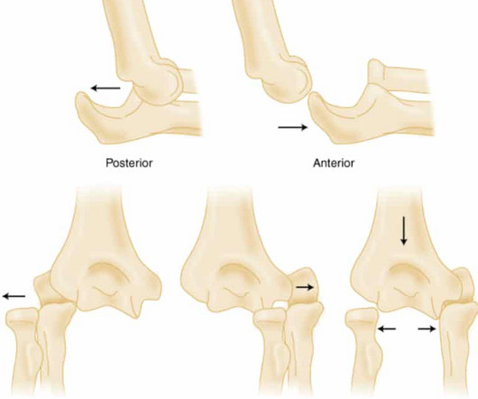
Elbow Dislocation Definition: Disarticulation of the proximal radius & ulna bones from the humerus Epidemiology: Incidence Second most common joint dislocation (after shoulder) in adults Most commonly dislocated joint in children Accounts for 10-25% of all injuries to the elbow ( Cohen 1998 ) Posterolateral is the most common type of dislocation (..)
Using a dedicated MRI system in the ED offers many potential benefits, from improving access to the modality for trauma patients to offering a more certain diagnosis than other imagingmodalities can, said presenter Catherine Mandel, MD, of the University of Melbourne in Australia. The bottom line?
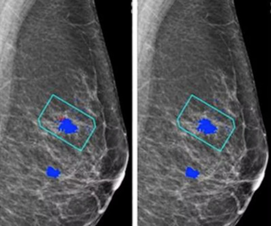
AI software based on deep-learning algorithms is showing promise, however, for helping to improve specificity in screening mammography and other breast imagingmodalities. Although mammogram is the most widely used screening modality, a known problem is that 9.5% Just over 40.5 million mammograms were performed in the U.S.
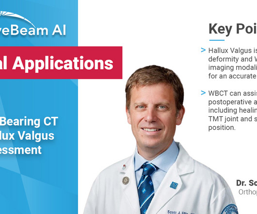
Key Points: Weight Bearing (WB) radiographs are limited by 1) their 2D nature and 2) knee positioning. Weight Bearing CT (WBCT) imaging provides three dimensional (3D) images of the knee compartment that could allow for earlier detection of osteoarthritis (OA). X-Ray (XR) beam angle differs by person and can be unreliable.
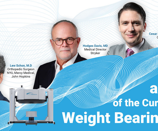
Key Points: The most important advantage of weight bearing CT (WBCT), which utilizes cone beam CT (CBCT) technology (a three-dimensional (3D) imagingmodality) is immediate access to 3D images, resulting in faster and better diagnostic capabilities.
The enduring shortage is affecting staffing for radiographers and radiologists; and all imagingmodalities. Marion Anderson, CRA, Diagnostic Imaging Manager at Karmanos Cancer Center in Detroit, Michigan reports that her facility is down two-thirds of staff for CT and mammography exams. Bureau of Labor Statistics.
If you work in radiology in a community healthcare facility, you may have low demand for pediatric medical imaging. I am passionate about improving the performance and interpretation of pediatric medical imaging. The balance of dose and image quality is even more important in pediatric medical imaging.
Kim Mason Kim Mason, an Audit and Research Radiographer for Mid Yorkshire Teaching Hospitals Trust, talks about their role as well as the value of radiographer engagement in research activities and how to get involved. Hi, I’m Kim and I am an alternative-styled, funky-haired, septum-pierced, disabled Audit and Research Radiographer.
This description highlights the significant advancements in medical imaging that are transforming healthcare. Advanced algorithms can analyze images more quickly and accurately, aiding in early diagnosis and treatment planning.
Reading Time: 4 minutes read Key takeaways from the 2023 RSNA Conference The topic of artificial intelligence technology trends in medical imaging was once again infused throughout sessions and on display in the exhibitor hall at the 2023 Radiological Society of North America (RSNA) scientific assembly and annual meeting.
This blog delves into the captivating realm of radiology and the intricate art of crafting diagnostic images with X-ray precision. From the subtlest nuances of bone structures to the shadowy landscapes of soft tissues, the precision of X-rays in creating images is akin to an artist’s brush painting upon a canvas.
Today, the same should be said of AI ROI image-guided technology and the radiation reduction and protection it provides. This is no less the case for interventional imaging. Interventional imaging extends the reach of human vision and enables greater patient care. That technology is now standard – as it should be.
A snapshot of the life of a radiology trainee… Clinical Radiology is a specialty that involves utilising imagingmodalities to help clinicians in the management of patients across all branches of medicine and surgery. It is true that a typical day as a radiology trainee involves performing and interpreting routine imaging.
CT Scan, PET CT Scan , PSMA PET CT Scan, Xray or MRI are common images that have to be interpreted & quickly delivered to the health care professionals who now demand quicker service. These new technological advancements have resulted in new imaging requirements that are increasingly complex.
Key Points: Weight bearing CT (WBCT) can detect signs of osteoarthritis (OA), such as osteophytes, subchondral cysts, and joint space narrowing better than radiographs. Emerging evidence suggests WBCT reveals meniscal extrusion not detected by MRI and WBCT arthrography visualizes meniscal tears.
Diagnosis While radiographs are typically sufficient to make the diagnosis, WBCT scans may be useful to plan surgical treatment. Accurately assess sesamoid position as plain radiographs cannot determine whether the sesamoids have been reduced within their grooves 5. Accurately assess healing in the 1st TMT joint 4.
After working for weeks in his lab experimenting on the production of ‘strange rays’, which he referred to as ‘X’, he asked his wife Anna Bertha to lend ‘a hand’, the left one to be precise, which he used to produce the first X-ray image. after seeing the image. (2) Photoprint from radiograph by W.K. Röntgen, 1895.
What are the important findings in each image. A: Coronal CT image demonstrates normal contour of the right globe (green arrow) and a shrunken left globe (orange arrow), which is suggestive of globe rupture. C: Sagittal CT image demonstrates normal contour in the right globe (green arrow). Imaging of orbital trauma.
There is no history or physical exam feature that rules out the disease Lactate elevation is a late finding in SBO. A normal lactate does not rule out the diagnosis Plain X-rays perform poorly in making or ruling out the diagnosis. 2.3 – 5.4) (-) LR: 0.18 (0.09 – 0.35) Ultrasound Findings Dilated loops of bowel (diameter > 2.5
Weight bearing CT (WBCT) imaging has fundamentally changed the evaluation and management of foot and ankle disorders such as hallux valgus, progressive collapsing foot disorder (PCFD), ankle instability, and traumatic deformities.
For dense-breasted patients requiring supplemental imaging, MRI remains a valuable option that is not limited by breast density and is shown to be more sensitive than mammography at finding breast cancer. vi Investigations continue of this newer imagingmodality, which has the potential to positively benefit patients with dense breasts.
In the world of orthopedic diagnostics, imaging plays a crucial role in identifying deformities and planning surgical interventions. Conventional radiographs and MRIs have been the standard, but they come with limitations when it comes to understanding the complex, three-dimensional structure of the human body. Why Choose WBCT?
Key Points: Imagingmodalities such as plain radiographs (X-Ray), computed tomography (CT), and magnetic resonance imaging (MRI), dont have the diagnostic accuracy needed to detect syndesmotic widening or subtle instability.
What if X-ray imaging, the most prevalent and accessible imagingmodality in the world, could provide the information needed for diagnosis? The study could lead to radiographs providing an early window into disease manifestations that are currently undetected, thus leading to an earlier diagnosis of IPF.
Newborns' livers can be affected by a variety of congenital and acquired diseases, and imaging plays an important role in the workup and management of these, according to a study published November 7 in RadioGraphics. Imaging has [a key] role in the workup and management of many neonatal hepatic abnormalities. msec (A), 5.36
However, despite frequent use, many emergency physicians are not familiar enough with stroke imaging to interpret images on their own. Acute stroke imaging is obtained in the emergency department for two purposes. 4] These findings are consistent with other studies and highlights the limitations of NCCT in acute stroke imaging.
We organize all of the trending information in your field so you don't have to. Join 5,000 users and stay up to date on the latest articles your peers are reading.
You know about us, now we want to get to know you!
Let's personalize your content
Let's get even more personalized
We recognize your account from another site in our network, please click 'Send Email' below to continue with verifying your account and setting a password.


Let's personalize your content