This site uses cookies to improve your experience. To help us insure we adhere to various privacy regulations, please select your country/region of residence. If you do not select a country, we will assume you are from the United States. Select your Cookie Settings or view our Privacy Policy and Terms of Use.
Cookie Settings
Cookies and similar technologies are used on this website for proper function of the website, for tracking performance analytics and for marketing purposes. We and some of our third-party providers may use cookie data for various purposes. Please review the cookie settings below and choose your preference.
Used for the proper function of the website
Used for monitoring website traffic and interactions
Cookie Settings
Cookies and similar technologies are used on this website for proper function of the website, for tracking performance analytics and for marketing purposes. We and some of our third-party providers may use cookie data for various purposes. Please review the cookie settings below and choose your preference.
Strictly Necessary: Used for the proper function of the website
Performance/Analytics: Used for monitoring website traffic and interactions
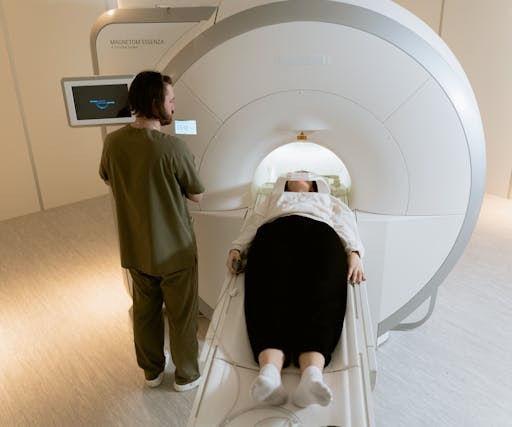
Using AI with mammography can help select women at high risk of breast cancer for supplemental MRI, according to research published February 4 in Radiology. Women at high risk of breast cancer, such as those with a personal history or family history of the disease or those with dense breasts, may be recommended for supplemental imaging.
Mammograms are a crucial diagnostic tool that helps doctors detect early signs of breast cancer and other breast-related issues. The truth is mammograms are generally safe when used properly, and the amount of radiation you’re exposed to is minimal. Radiation exposure is controlled and minimized to ensure patient safety.
An act making mammograms more accessible in Illinois is now in lawmakers’ hands. Coverage for molecular breast imaging will be required and, in those cases where it is not already covered, breast MRI will also be covered. HB 4180 is sponsored by Reps. Nabeela Syed (D-51st District), Michael J.
AI software based on deep-learning algorithms is showing promise, however, for helping to improve specificity in screening mammography and other breast imaging modalities. million mammograms were performed in the U.S. Although mammogram is the most widely used screening modality, a known problem is that 9.5% Just over 40.5
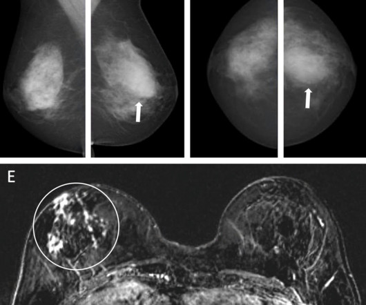
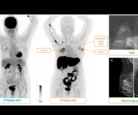
Nuclear medicine physicians analyzed both sets of images and determined disease stages, with final stages verified via biopsy. (B) B) F-18 FDG-PET maximum intensity projection, F-18 FES-PET maximum intensity projection, breast MRI scan, and mammogram in an 81-year-old female participant who presented with a tumor in the right breast.
Abbreviated MRI may be suitable for screening women with dense breasts, a study published May 22 in the American Journal of Roentgenology found. Radiologists and ordering clinicians, alike, may use these results as evidence of the value of abbreviated MRI beyond the first round of screening in this population," Edmonds told AuntMinnie.com.
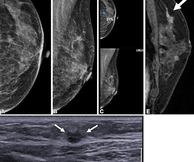
Postoperative MRI surveillance appears to lower the odds of advanced second breast cancer in women with a personal history of the disease, researchers have reported. "In Images in a 40-year-old woman who underwent breast-conserving surgery for left breast cancer and a surveillance breast MRI examination 25 months after surgery. (A)
As the initial COVID-19 vaccines became available in the second year of the pandemic, reports emerged of imaging findings associated with vaccination. Vaccinated patients could demonstrate imaging results of how vaccines could impact imaging results. Images and captions courtesy of the American Roentgen Ray Society.
Breast density can often obscure lesions on conventional x-ray mammography, and so other screening modalities such as MRI or ultrasound are often recommended for follow-up. CEM is faster and less costly than MRI and can often be used as a follow-up to an abnormal screening mammogram when it is clinically appropriate.
Low-dose positron emission mammography (PEM) can detect invasive breast cancer in a feasible manner, according to research published February 9 in Radiology: Imaging Cancer. This study underscores the potential of this low-dose PEM system as a promising imaging tool in breast cancer diagnosis,” the Freitas team wrote.
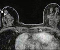
However, the researchers noted a lack of data on interpreting surveillance mammograms in women with a personal history of breast cancer. Images depict a 65-year-old patient with contralateral second breast cancer 6.3 A) Left craniocaudal and (B) mediolateral oblique mammograms assessed as benign. (C)
A radiologist’s perception when viewing a complex MR image may be akin to a Major League Baseball (MLB) batter reading the stitches on a fastball, according to researchers exploring exactly how diagnostic interpretations are made. The Perception Lab is a "pop-up" version of an academic research lab focused on medical image perception.
Komen has urged quick passage in Arizona of legislation that would eliminate patient out-of-pocket costs for diagnostic and supplemental breast imaging. House Bill 2411 was introduced in the state by Representative David Cook (R-Globe) and includes eliminating costs for patients for MRI, ultrasound, and diagnostic mammograms.
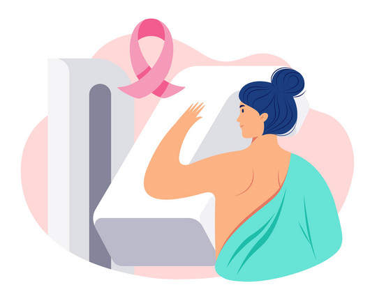
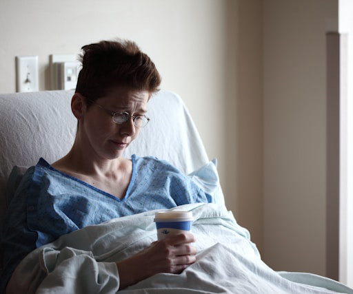
Amidst the battle against this disease, screening mammograms emerge as a crucial tool in early detection and effective treatment. In this blog, we delve into the significance of screening mammograms, their procedure, their benefits, and why they are essential for women’s health. What is a Screening Mammogram?
Cryoablation uses imaging guidance typically with ultrasound or CT to locate tumors. Follow-up imaging was performed after the procedure by mammogram, ultrasound, or in some cases contrast-enhanced mammogram or MRI, based on patient eligibility and preference.
Previous studies have demonstrated that dense breast tissue masks breast cancers on mammography, and that supplemental imaging such as ultrasound and MRI confirms suspicious findings within dense tissue. Kerlikowske has studied breast density on imaging. The law goes into effect September 10, 2024. “The
An AI-based method can outperform models based just on breast density analysis for identifying patients who would benefit from supplemental imaging, according to research published April 9 in Radiology. What’s more, the AI score could potentially have saved more than twice the number of years of life than relying on density measurements. “In
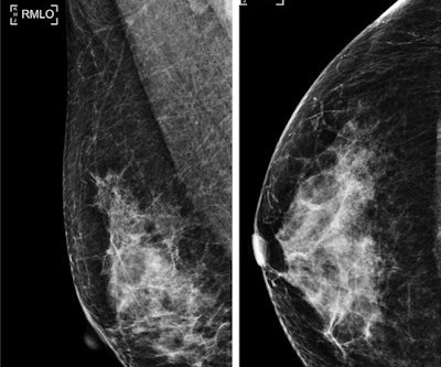
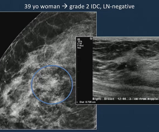
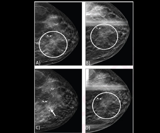
The retrospective study included data collected between 2007 and 2019 from 4,150 women in this age range who underwent 4,448 screening mammograms. Images of a 39-year-old woman shows a grade 2 invasive ductal carcinoma on mammography, confirmed by supplementary ultrasound.
Breast imaging organizations and political bodies are criticizing updated guidelines on breast cancer screening by the Canadian Task Force on Preventive Health Care (CTFPHC). Additionally, the guidelines do not recommend supplemental imaging in women with dense breasts or women with a personal family history of breast cancer.
Breast imaging experts are criticizing a recent study that questioned whethe. Read more on AuntMinnie.com Related Reading: Breast MRI uptake is low among women at high breast cancer risk Online self-scheduling use in mammography increases Do cancer screening exams really extend patients' lives?
Some factors tied to first breast cancers can have a negative impact on screening mammography's ability to find future cancers, according to research published October 5 in Clinical Imaging. Also, mammogram-occult primary breast cancer is the most important risk factor for a contralateral mammogram-occult second breast cancer, the team found.
Contrast-enhanced mammography (CEM) has tradeoffs when it comes to imaging women with extremely dense breasts, according to research published October 1 in Radiology. This problem has led to researchers exploring different methods for better accuracy in imaging these women. Image courtesy of the RSNA. 0.003 Specificity 96.2%
The reduction for the bilateral mammogram 77066 was 1.36%, reflecting an increase in RVU valuation that somewhat offsets the conversion factor cut. For example, an imaging center with all the listed modalities except for PET would see a decrease of 2.6% rather than 2.5%
A deep-learning model based on mammographic images can determine tumor staging and sentinel lymph node metastasis in breast cancer patients, a study published April 15 in Informatics in Medicine Unlocked found. The researchers highlighted that CNNs require little preprocessing compared with other image classification algorithms.
A team led by Joao Horvat, MD, from the Memorial Sloan Kettering Cancer Center in New York found that CEM depicted 90% of breast cancers compared with 10% on low-energy mammograms alone and 50% on low-energymammogramswith whole-breast ultrasound. Horvat and co-authors investigated whether the same trend goes for CEM.
Dense Breast Info (DBI) has issued a statement of support for “Find It Early Act” HR 3086, a bill that addresses insurance barriers to breast cancer screening beyond an initial mammogram. Food and Drug Administration (FDA) will require that women be informed after their mammograms whether their breasts are “dense” or “not dense.”
Food and Drug Administration (FDA) has said CT is still the preferred imaging modality for patients with medical devices. However, "for imaging procedures specifically, CT continues to be the preferred tomographic imaging technology for people with implantable or wearable medical devices. Read the FDA's full guidance here.
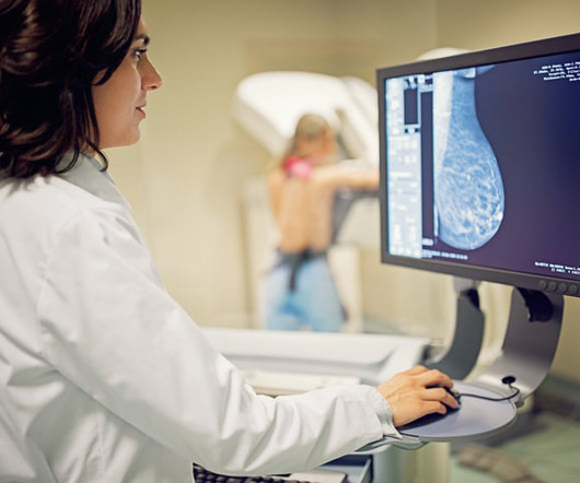
Advanced imaging technologies continue to play a crucial role in detecting cancers before they progress, giving patients the best chance for successful treatment. The Importance of Early Detection Through Imaging Early detection of cancer through imaging allows for interventions at stages when treatment is most effective.
Imaging surveillance may be a safe alternative to surgical excision for architectural distortions detected by digital breast tomosynthesis (DBT) alone, suggest findings published March 12 in the American Journal of Roentgenology. C) Post-biopsy mammogram shows cylinder clip (arrow) in appropriate position.
The American Cancer Society recommends starting annual mammogram screenings at age 40. In some instances, other imaging exams may be requested for many reasons. Some of these reasons include: incomplete information, something is found that needs further investigation, other image angles are needed to get a better view, etc.
This breast surgery is often performed long before a woman needs her first mammogram , which means that the topic of breast implants’ effects on necessary screenings is rarely discussed before breast augmentation is performed. How often do I need a mammogram? Mammograms should be scheduled every 1 to 2 years until at least age 75.
Why Breast Density Matters in Cancer Screening Dense breast tissue affects screening in two key ways: Reduced Visibility : Dense tissue appears white on mammograms, as do tumors, making it harder to detect abnormalities. Inter-radiologist Variation : Assessments can vary up to 33% 1 when different radiologists interpret the same mammograms.
IMI joins SLHP, providing quality and affordable medical imaging to the Treasure Valley Healthcare Community {Boise, Idaho, February 15, 2024} SLHP members now have more options when it comes to accessing medical imaging within the Treasure Valley. Luke’s Health Partners.
For dense-breasted patients requiring supplemental imaging, MRI remains a valuable option that is not limited by breast density and is shown to be more sensitive than mammography at finding breast cancer. Ultrasound rounds out the radiologist’s toolkit for supplemental imaging of women with dense breasts.
If you’re in your 30’s, you may be wondering if it’s time to add a yearly mammogram to your healthcare routine. When Does the American Cancer Society Recommend Starting Mammograms? Continue to get mammograms if you’re in good health and expect to live an additional 10 years. Should I Get a Mammogram in My 20s or 30s?
High-Risk Screening Program Midstate Radiology Associates, one of the first Radiology providers in the United States to offer a high-risk screening program within all Womens Imaging locations, is a proud partner of CARE, a secure application to collect and analyze patients medical information and family history.
Capitol Imaging Services has served tens of thousands of women with our women’s health examinations in screening mammography, diagnostic mammography, ultrasound, bone density studies, breast MRI and breast biopsy. For screening mammograms, we ask for about 20 minutes of a woman’s time. Convenient.
In Michigan, a patient recently reported waiting over 80 days for imaging results. Another waited three months for mammogram findings. Theyre part of a larger trend, driven by a persistent imbalance between the number of radiologists available and the ever-growing demand for diagnostic imaging. These delays arent isolated.
While this is an improvement over the prior recommendation to begin screening at age 50, it falls short of the current recommendation by the Society of Breast Imaging and the American College of Radiology to obtain annual mammograms beginning at age 40. Concern for radiation risk due to annual screening is really of no concern!
Further, the task force incorrectly concludes there is “inadequate” evidence to support adding screening MRI (or ultrasound if MRI is not possible) after a mammogram for many women with dense breasts. In women with the densest breasts, about 40% of cancers are missed on a mammogram.
The earlier breast cancer is detected through diagnostic imaging, the better chance there is for successful treatment with surgery, radiation therapy, or chemotherapy. Consistent mammography and diagnostic imaging appointments can help stop this number from continuing to rise. this year alone. this year alone.
milla1cf Wed, 07/05/2023 - 19:42 uly 5, 2023 — A new study by investigators from Mass General Brigham has found that artificial intelligence ( AI ) language models like ChatGPT can accurately identify appropriate imaging services for two important clinical presentations: breast cancer screening and breast pain. ChatGPT 4 outperformed 3.5,
We organize all of the trending information in your field so you don't have to. Join 5,000 users and stay up to date on the latest articles your peers are reading.
You know about us, now we want to get to know you!
Let's personalize your content
Let's get even more personalized
We recognize your account from another site in our network, please click 'Send Email' below to continue with verifying your account and setting a password.


Let's personalize your content