This site uses cookies to improve your experience. To help us insure we adhere to various privacy regulations, please select your country/region of residence. If you do not select a country, we will assume you are from the United States. Select your Cookie Settings or view our Privacy Policy and Terms of Use.
Cookie Settings
Cookies and similar technologies are used on this website for proper function of the website, for tracking performance analytics and for marketing purposes. We and some of our third-party providers may use cookie data for various purposes. Please review the cookie settings below and choose your preference.
Used for the proper function of the website
Used for monitoring website traffic and interactions
Cookie Settings
Cookies and similar technologies are used on this website for proper function of the website, for tracking performance analytics and for marketing purposes. We and some of our third-party providers may use cookie data for various purposes. Please review the cookie settings below and choose your preference.
Strictly Necessary: Used for the proper function of the website
Performance/Analytics: Used for monitoring website traffic and interactions
CHICAGO -- When AI in mammography is coupled with a safeguard radiologist review, women are more likely to pay for breast cancer screening mammography if the AI will boost exam results, research presented December 5 at the RSNA meeting suggests. Investigators explored the impact of adding AI to mammogram screening exams. versus 4.6%.
Viewing patients' priors consistently improves readers' performances, regardless of experience level, specialization or the volume of screening mammograms they are accustomed to reading.
Women in racial and ethnic minority backgrounds are less likely to be provided same-day diagnostic breast imaging services, despite such services being available, according to research published February 18 in Radiology. Additional imaging and possibly image-guided biopsy are recommended for women who have an abnormal screening mammogram.
A radiologist’s perception when viewing a complex MR image may be akin to a Major League Baseball (MLB) batter reading the stitches on a fastball, according to researchers exploring exactly how diagnostic interpretations are made. The Perception Lab is a "pop-up" version of an academic research lab focused on medical image perception.
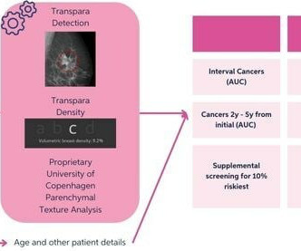
Women at high risk of breast cancer, such as those with a personal history or family history of the disease or those with dense breasts, may be recommended for supplemental imaging. ScreenPoint Medical ) for breast cancer detection on 2D mammograms to triage women with an assumed intermediate breast cancer risk for supplemental MRI.
Standalone AI can significantly outperform radiologists' sensitivity in reading digital mammograms and has shown potential in DBT exams as well, but experts are not yet ready to hand over the reins.
An act making mammograms more accessible in Illinois is now in lawmakers’ hands. Coverage for molecular breast imaging will be required and, in those cases where it is not already covered, breast MRI will also be covered. HB 4180 is sponsored by Reps. Nabeela Syed (D-51st District), Michael J.
Radiologists interpreting screening mammograms may be especially susceptible to falling victim to automation bias, as these exams are repetitive in nature.
AI software based on deep-learning algorithms is showing promise, however, for helping to improve specificity in screening mammography and other breast imaging modalities. million mammograms were performed in the U.S. Although mammogram is the most widely used screening modality, a known problem is that 9.5% Just over 40.5
When it comes to working up palpable breast abnormalities with contrast-enhanced mammography (CEM), recombined images better depict the lesion than low-energy ones, research published November 8 in Academic Radiology has found. The investigators used pathology or one-year follow-up imaging as the reference standard.
Breast radiologists may want to consider the physical surroundings of mammography rooms for optimal image quality, according to a quality control study published November 15 in Radiography. However, white wall color around monitors and high ambient light have negative impacts on image evaluation.
While the pandemic affected medical operations across the country, the experts said that radiologists developed and honed their sense of resiliency as imaging was placed on the front lines. Medical imaging played a significant role in the early days of the pandemic when it hit its initial peak in April 2020.
Maybe you recently decided to try the best 3D mammogram experience in El Paso, have recently moved, changed doctors, or acquired new insurance, and are now going to our imaging center for your annual mammogram. So, why are these prior images so important? In this case, the radiologist may recommend a diagnostic mammogram.
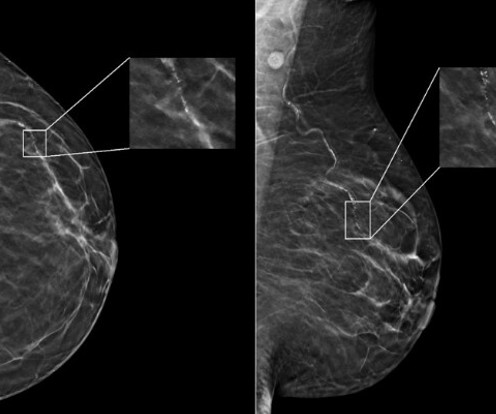
Screening mammography exams arranged from low to high breast density can boost radiologists' interpretation performance, suggest findings published on October 8 in Radiology. This idea stems from previous studies' findings that between 20% and 50% of interval and screen-detected breast cancers were visible on prior screening mammograms.
Radiologists interpreting screening mammograms may be especially susceptible to falling victim to automation bias, as these exams are repetitive in nature.
This goes for women in all age groups and women who are Asian, Black, Hispanic, and Native American, according to the report written by Edward Hendrick, PhD, from the University of Colorado in Aurora and Debra Monticciolo, MD, from the Foundation for Imaging Research and Education in Temple, TX. They added that the U.S.
As the initial COVID-19 vaccines became available in the second year of the pandemic, reports emerged of imaging findings associated with vaccination. Vaccinated patients could demonstrate imaging results of how vaccines could impact imaging results. Images and captions courtesy of the American Roentgen Ray Society.
Imaging societies and cancer screening advocates continue to criticize a recent federal ruling that challenges no-cost preventive health services. Becerra, imaging society experts said it could open the door for future legal challenges to such services. Xavier Becerra et al. While the ruling only impacts parties in Braidwood v.
In breast imaging, AI has shown its potential as a clinical assistant to interpreting radiologists. To do so, three radiologists annotated a dataset of mammograms using histology-based ground truth. These networks were also validated and tested using an annotated dataset of 1,000 patients and 1,986 mammograms.
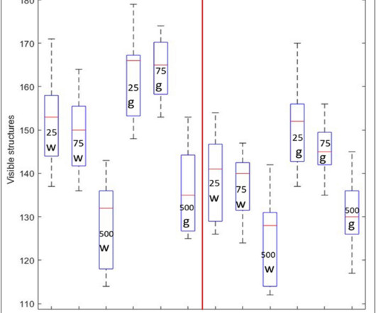
Curve-shaped breast compression paddles may lead to some pain relief, but they also reduce image quality on mammograms, suggest findings published August 20 in Radiology. But the paddles also decreased image contrast and the visibility of structures. who were screened at three sites in the Netherlands.
With AI-mediated cyberattacks on the rise, radiologists can find it challenging to balance prompt patient access to diagnostic imaging while safeguarding sensitive healthcare data. With growing radiology practices and outpatient imaging facilities, we will see an increase in outsourced diagnostic imaging. Dhaval Shah.
milla1cf Mon, 05/22/2023 - 13:25 May 22, 2023 — Incorrect advice by an AI-based decision support system could seriously impair the performance of radiologists at every level of expertise when reading mammograms, according to a new study published in Radiology , a journal of the Radiological Society of North America ( RSNA ).
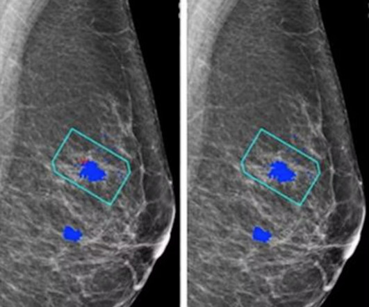
Radiology departments continue to have interest in implementing AI into their workflows, with the technology being used to manage workloads among radiologists. iCAD) generated case scores (malignancy certainty) and risk scores (one-year subsequent malignancy risk) for each mammogram. The FDA-approved algorithm (ProFound AI 3.0,
The finding suggests the technique may provide a new treatment path for women who are not candidates for lumpectomy, or surgical removal, noted Yolanda Bryce, MD, an interventional radiologist at Memorial Sloan Kettering Cancer Center in New York City, and senior author of the study.
A deep-learning algorithm can rule out the presence of breast cancer on screening mammograms, improving specificity and yielding significant workflow and downstream savings, according to research published April 10 in Radiology. dataset 1: 143,593 mammograms interpreted by 11 breast radiologists from 2008 to 2017 U.S.
CureMetrix plans to roll out its cmAngio AI software to SimonMed Imaging's network of U.S. Food and Drug Administration (FDA), CureMetrix noted, adding that the software will be used to read mammograms and detect and localize breast arterial calcifications. SimonMed Imaging operates in 10 states. physician radiology practices.
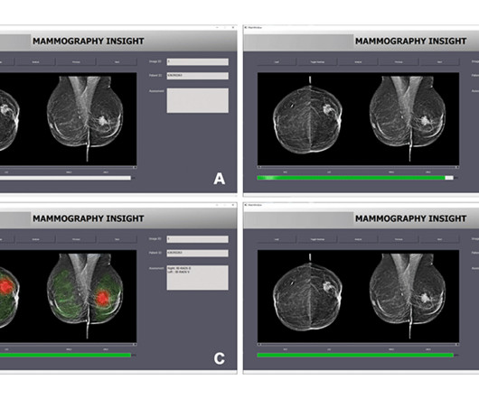
christine.book Wed, 11/22/2023 - 12:11 November 22, 2023 — ScreenPoint Medical has announced that its Transpara breast AI has surpassed 5 million mammograms, including over 1 million Tomosynthesis (3D) exams analyzed in support of radiologists reading mammography exams. Gail, TC8). "As
A recent Australian study criticizing narratives surrounding breast density notification has drawn mixed reactions from radiologists and advocates of breast cancer screening. Radiologists have studied and continue to research how breast density plays into breast cancer risk. Kerlikowske has studied breast density on imaging.
Tucked away beneath all of the symbolism and public events, is the quiet experience of the mammogram and the radiology technology that makes it possible. Why are mammograms at the center of this public health battle? The advent of mammography remains the go-to for breast imaging. Why is a Mammogram Important?
ChatGPT demonstrates modest accuracy when assigning BI-RADS scores for mammograms and breast ultrasound exams, according to research published October 30 in Clinical Imaging. Previous reports suggest that large language models can correctly recommend appropriate imaging modalities for patients based on their clinical presentation.
We're excited to introduce advanced workstation features for our flagship solution, ProFound Detection, aimed at further improving and facilitating radiologists' interpretation of mammograms within their workstation,” said Dana Brown , President and CEO of iCAD.
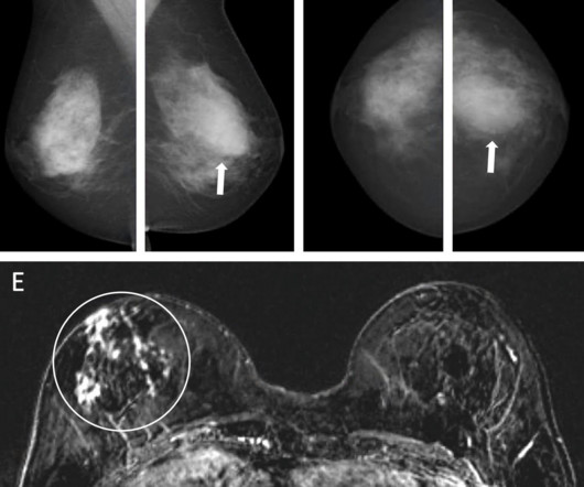
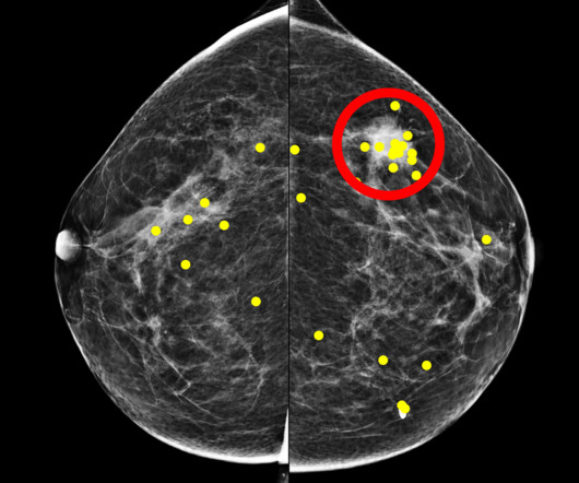
Healthcare disparities continue to plague medical imaging, but there are concrete measures radiologists can take to mitigate them, according to a paper published on October 12 in RadioGraphics. B) A craniocaudal magnified mammogram more clearly shows the irregular mass and pleomorphic calcifications in the medial breast. (C)
The Benefits of Regular Mammograms for Women Mammography plays a crucial role in breast cancer prevention by contributing to early detection, which is one of the most effective ways to reduce mortality and improve treatment outcomes. Schedule your mammogram appointment today at (915) 225-2480 ! They are considered precancerous changes.
Low-dose positron emission mammography (PEM) can detect invasive breast cancer in a feasible manner, according to research published February 9 in Radiology: Imaging Cancer. This study underscores the potential of this low-dose PEM system as a promising imaging tool in breast cancer diagnosis,” the Freitas team wrote.
As a tenant in the Milford Walmart Supercenter, MammogramNow delivers essential breast screening services, provided by a team of board-certified radiologists and trained technologists from RadNet’s Delaware Imaging Network.
Breast density assessment is important not only for the patients health, but it also has reimbursement implications for radiologists. Considerations for subsequent imaging Patients identified as having dense breasts are recommended for additional imaging using MRI, ultrasound, or contrast-enhanced mammography (CEM).
An AI-based method can outperform models based just on breast density analysis for identifying patients who would benefit from supplemental imaging, according to research published April 9 in Radiology. What’s more, the AI score could potentially have saved more than twice the number of years of life than relying on density measurements. “In
Amidst the battle against this disease, screening mammograms emerge as a crucial tool in early detection and effective treatment. In this blog, we delve into the significance of screening mammograms, their procedure, their benefits, and why they are essential for women’s health. What is a Screening Mammogram?
christine.book Tue, 05/21/2024 - 10:36 May 21, 2024 — According to a newly-published study of nearly 5,000 screening mammograms interpreted by an FDA-approved AI algorithm, patient characteristics such as race and age influenced false positive results. Duke Health , breast radiologist and assistant professor at Duke University in Durham, NC.
Incorrect advice by an AI-based decision support system could seriously impair the performance of radiologists at every level of expertise when reading mammograms, according to a new study published in Radiology.
The challenge aims to develop an architecture capable of automatically estimating the breast percentage density from mammograms. These tools also require an experienced radiologist to adjust the segmentation threshold for dense tissue within the breast area. High breast tissue density is a significant risk factor for breast cancer.
We organize all of the trending information in your field so you don't have to. Join 5,000 users and stay up to date on the latest articles your peers are reading.
You know about us, now we want to get to know you!
Let's personalize your content
Let's get even more personalized
We recognize your account from another site in our network, please click 'Send Email' below to continue with verifying your account and setting a password.


Let's personalize your content