This site uses cookies to improve your experience. To help us insure we adhere to various privacy regulations, please select your country/region of residence. If you do not select a country, we will assume you are from the United States. Select your Cookie Settings or view our Privacy Policy and Terms of Use.
Cookie Settings
Cookies and similar technologies are used on this website for proper function of the website, for tracking performance analytics and for marketing purposes. We and some of our third-party providers may use cookie data for various purposes. Please review the cookie settings below and choose your preference.
Used for the proper function of the website
Used for monitoring website traffic and interactions
Cookie Settings
Cookies and similar technologies are used on this website for proper function of the website, for tracking performance analytics and for marketing purposes. We and some of our third-party providers may use cookie data for various purposes. Please review the cookie settings below and choose your preference.
Strictly Necessary: Used for the proper function of the website
Performance/Analytics: Used for monitoring website traffic and interactions
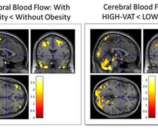
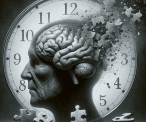
CHICAGO -- Characterizing an individual's type of body fat using body MRI can help predict Alzheimer's disease risk up to 20 years before symptoms manifest, according to research results presented December 2 at the RSNA meeting. Images courtesy of the RSNA.
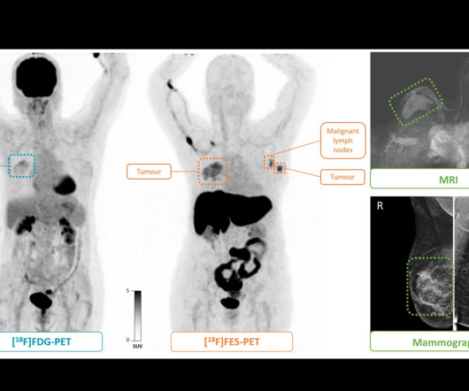
Between December 2018 and January 2021, the group enrolled 41 female participants (median age, 56 years) who underwent both PETscans. Nuclear medicine physicians analyzed both sets of images and determined disease stages, with final stages verified via biopsy. (B) All lesions had also been identified at mammography and MRI.
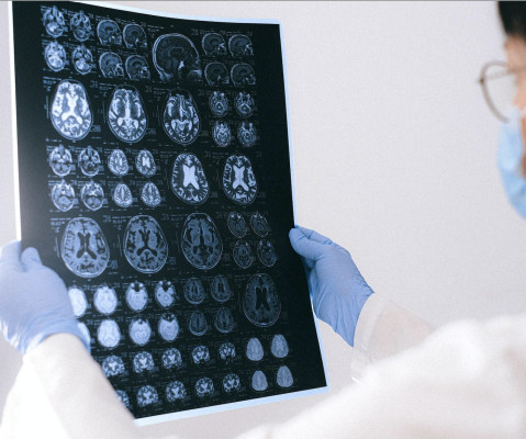
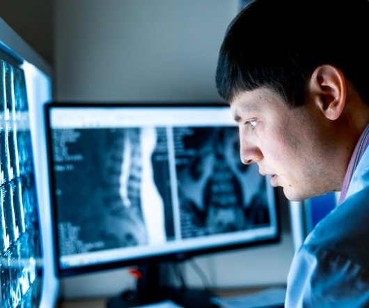
The way physicians identify illness is changing due to advances in medical imaging, which make early diagnosis quicker, more precise, and less invasive. These technologies, ranging from high-resolution MRIs to state-of-the-art CT scans, can give doctors the ability to spot possible health problems before symptoms even show up.
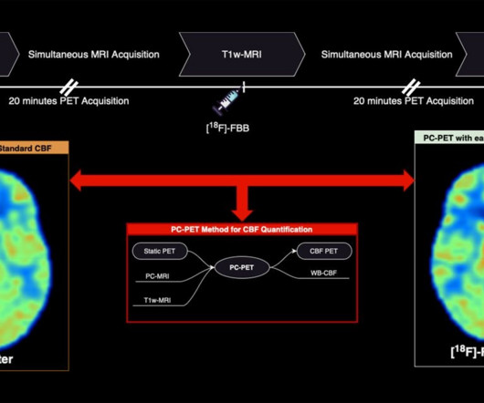
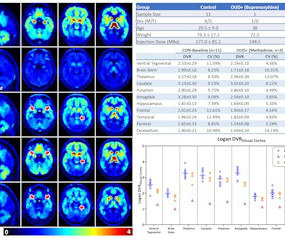
A PET radiotracer for diagnosing Alzheimer’s disease may also be used to measure vascular brain changes in patients during PET/MRIscans, according to a study published December 7 in the Journal of Nuclear Medicine. Image courtesy of the Journal of Nuclear Medicine. The researchers enrolled 20 participants.
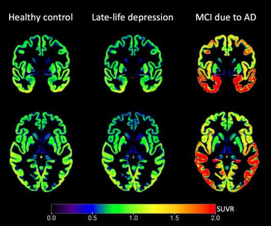
A PET/MRI study has provided insights into the neurobiology of late-life depression, with researchers reporting that tau protein – a key protein involved in Alzheimer’s disease – does not appear to be involved in the condition. Image and caption courtesy of the American Journal of Geriatric Psychiatry.
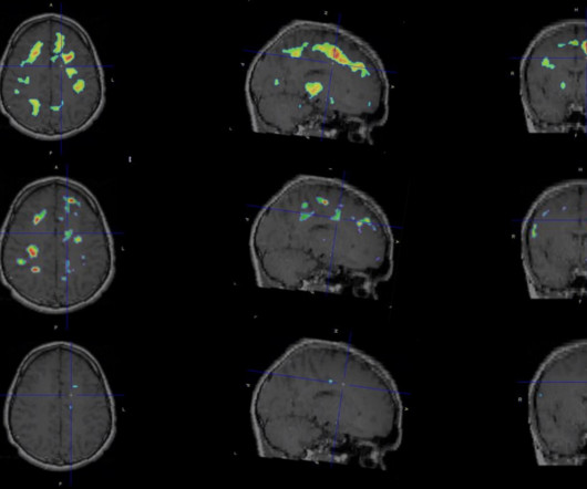
An AI algorithm designed for brain PETimaging found a glioblastoma in a patient that had gone undetected by physicians, according to a case reported February 15 in the Journal of Nuclear Medicine. Baseline scan (A), segmentation results (B), and follow-up scan (C) from patient with molecular glioblastoma.
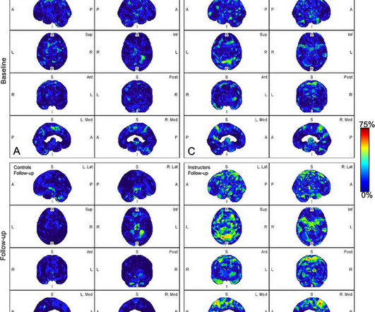
PET brain scans show persistent brain inflammation in patients with multiple sclerosis (MS), despite being treated with high-efficacy disease-modifying therapies, according to a recent study by researchers in Boston. The researchers performed F-18 PBR06 PETscans on 22 patients with MS and eight healthy controls.
PETimaging shows different brain activity in people taking methadone or buprenorphine for opioid use disorder (OUD), according to a presentation at the recent Society of Nuclear Medicine and Molecular Imaging annual meeting. Image and caption courtesy of Jacob Dubroff, MD, PhD.
FDG-PETimaging shows promise for use as a diagnostic criterion for neurosarcoidosis, with a recent case series illustrating the approach was effective when gold-standard approaches were not, according to a group of researchers in Berlin. “To In all three cases, gadolinium-enhanced MRIscans did not show abnormalities.
The following is the list of candidates for the 2024 edition of the Minnies, AuntMinnie.com 's campaign to recognize the best and brightest in medical imaging. Image from Martin W. tesla brain MRIscans. Image from Denis Le Bihan, PhD, of the NeuroSpin research facility, et al. AI detects cancer on breast MRI.
A group in England has established PETimaging as a new approach for studying gait – an excellent indicator of physical, emotional, and mental health, according to a study published February 6 in NeuroImage. F-18 FDG-PETscans allow clinicians to measure the brain's energy demands based on glucose metabolism.
According to clinical trials, amyloid PETscans showed that donanemab reduced amyloid plaques by up to 84% after 18 months of treatment. Safety warnings include that the drug can cause amyloid-related imaging abnormalities, which are detected by MRI and present as temporary swelling in an area or areas of the brain.
The team has worked for the past eight years on developing an approach of imaging cells called microglia. Microglia are immune cells in the brain that are thought to have a role in MS disease progression but cannot be seen by a routine MRI. The team developed a technique called F18 PBR 06 PETimaging.
The team investigated any associations between brain MRI volumes -- as well as amyloid and tau uptake on PETscans -- with body mass index (BMI), obesity, insulin resistance, and abdominal fatty tissue in cognitively healthy midlife individuals. Image courtesy of the RSNA. The green colors are the normal white matter.
PETimaging using a newly developed radiotracer has identified different patterns of brain tau pathology over time in early-onset versus late-onset Alzheimer’s disease patients, according to a study published February 1 in the Journal of Nuclear Medicine. in 15 patients with negative amyloid PETscans; 1.18 in
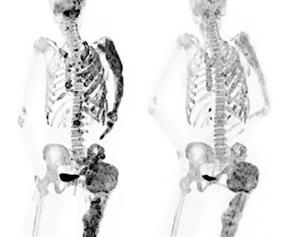
Read more on AuntMinnie.com Related Reading: NaF-PET shows bone formation in psoriatic arthritis patients PET/MRI provides new insights into knee osteoarthritis NaF-PET reveals aortic wall injuries NaF-PETscans reveal plaque -- and possible risk of stroke Can deep learning monitor lesions on F-18 NaF PET/CT?
iPET is AI-based software that denoises, sharpens organ boundaries, and improves resolution in PET and SPECT scans. Scans processed with the algorithm can also provide additional details by using an overlay of an MRI or CT image of the same region, the company said.
milla1cf Thu, 07/27/2023 - 23:20 July 27, 2023 — Scientists have found a new use for copper in magnetic resonance imaging (MRI) contrast agent design, that could help to create better images which help doctors diagnose patients’ conditions more easily and safely.
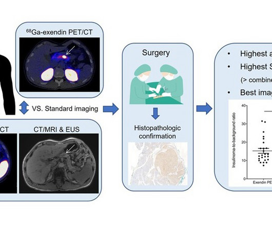
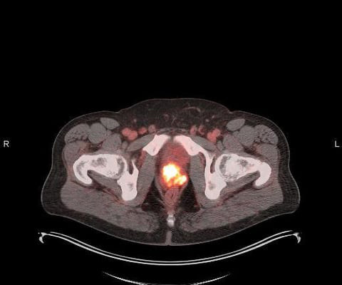
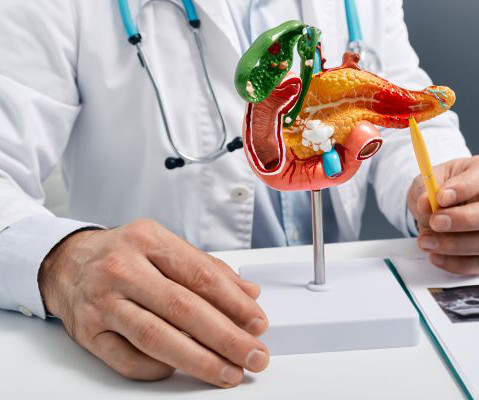
A new PETscan has shown promise in reliably detecting benign tumors in the pancreas that cannot be detected by various imaging techniques such as CT, MRI, and PETscans.
A new PETscan has shown promise in reliably detecting benign tumors in the pancreas that cannot be detected by various imaging techniques such as CT, MRI, and PETscans.
The promise of imaging One challenge is that advances in Alzheimer’s care can only help people if they are reaching the right patients at the right time in their patient journey. Reimbursement barriers Unfortunately, until recently, there were significant barriers to receiving amyloid PETimaging in some regions.
Still, these studies have included small samples and have not focused on developing an MRI biomarker. LEADS participants receive a standard clinical evaluation and Magnetic Resonance Imaging ( MRI ) scanning as well as amyloid and tau PETscanning and fluid biomarker assessments on an annual basis.
announced a research collaboration agreement with the Alzheimer’s Disease Neuroimaging Initiative 4, ADNI4, on the use of Meilleur’s [F-18]NAV-4694, an investigational imaging agent, in Positron Emission Tomography (PET) scans to assess the status amyloid plaque in the brain.
The company has been developing human digital twin technology at the organ and lesion level, as well as foundational AI models designed to extract actionable insights from MRI, CT, and PETscans. through the CE and UKCA marks, some of which are 510(k) cleared by the U.S.
As technological capabilities advance across all medical disciplines, so to do the imaging needs that often come with adequate diagnostic capabilities. The answer to many of these questions could be just one—MRI. MRIscans are also a common follow-up procedure used to investigate findings of other imaging techniques further.
Medical imaging has evolved over centuries, starting with X-rays in 1895, progressing to CT, MRI, and PETscans. The post Revolutionising Healthcare: A Historical Perspective on Medical Imaging appeared first on Open Medscience.
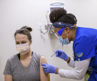
This can look like a clinically significant finding on cancer imaging, including chest CTs, PETscans, mammography, and breast MRI. A small but significant number of people experience swollen lymph nodes as a side effect of receiving a COVID vaccine. … Continue reading →
Non-invasive positron emission tomography , or PET, imaging could be used to identify early-stage amyloid-beta accumulation in individuals or professions exposed to traumatic brain injury such as military personnel, police officers, firefighters, football players, etc.,” The control participants were evaluated at similar time points.
milla1cf Tue, 05/30/2023 - 19:49 May 30, 2023 — Blue Earth Diagnostics , a Bracco company and recognized leader in the development and commercialization of innovative PET radiopharmaceuticals , today announced U.S. It is the first and only FDA-approved, PSMA-targeted imaging agent developed with proprietary radiohybrid (rh) technology.
As we bid adieu to the final moments of 2023, it’s a great time to reflect on advancements and studies that have redefined the world of imaging this year. In this article, we’ll delve into the hottest news and breakthroughs in imaging, highlighting the remarkable strides that have made the headlines.
While it forms in the tissues surrounding your body’s internal organs, location influences the symptoms and subsequent diagnostic procedures, which include imaging and imaging-guided biopsy. Imaging for Mesothelioma Medical imaging serves several purposes.
There’s a strong and innovative imaging department here, which will allow us to use MRI and sophisticated PETscanning to guide treatments, as well as outstanding urology and medical oncology departments, to provide the most comprehensive and multidisciplinary treatment recommendations.”
Brain Tumor Spotted on PETImaging An AI algorithm named “JuST_BrainPET” identified a glioblastoma in a patient that had been missed by physicians. The algorithm automatically segments metabolic tumor volume from healthy tissue on brain PETimaging.
In honor of Sarcoma Awareness Month, this blog post sheds light on these conditions and delves into the role of cutting-edge imaging technologies in advancing our understanding and management of sarcoma and bone cancer. Discuss the limitations of conventional imaging and the need for more advanced technologies for precise diagnosis.
Medical imaging crucially enhances oncology, aiding early cancer detection and effective treatment planning. The post The Transformative Impact of Medical Imaging in Oncology appeared first on Open Medscience.
The post Sustainable Imaging: Pioneering a Greener Future in Radiology appeared first on Open MedScience. Green radiology promotes sustainability by reducing energy use, managing waste, and integrating renewable resources effectively.
To confirm a diagnosis, a patient will have a biopsy, imaging and blood tests. CT Scans A computed tomography (CT) scan is useful for visualizing and creating a 3D representation of internal organs. Through the multidirectional images, doctors can identify the presence of a tumor and assess if the condition has spread.
Advances in medical imaging technology have significantly improved diagnostic accuracy, enabling earlier detection and more personalised treatments. The post Advances in Medical Imaging Technology: Unlocking the Mysteries of the Human Body appeared first on Open MedScience.
PACS – Picture Archiving and Communication System; a system involved in acquiring the medical images, transmission, viewing, storage, and retrieval of same images. The fundamental parts of PACS are – imaging acquisition, display workstations, archive servers. of imaging modalities, storage space, no.
Diagnosis often depends on the interpretation of tests like of X-Rays, CT Scan, MRI, PET CT Scan , etc. Digital Imaging and Communications in Medicine (DICOM) is used to transmit and store data. If there is a small community facility it can save costs by outsourcing the imaging and diagnosis.
How Much Does a PET/CT cost? A lot can go through your mind when your physician orders a PET/CT for you. Along with the normal anxiety associated with needing a medical imaging study, worrying about your out-of-pocket expense is a reasonable concern. But, first, let’s understand what a PET/CT is and why it is used.
Diagnostic imaging in motor neurone disease (MND) is crucial for early detection, disease monitoring, and differentiating from other conditions. The post Motor Neurone Disease: Diagnosis and Future Research Insights appeared first on Open MedScience.
PETscans have revealed for the first time what may be a key molecular driver of stress and addiction in people with alcohol use disorder (AUD), according to a study published January 21 in Neurobiology of Stress. Image courtesy of Neurobiology of Stress. According to the findings, PETscans revealed 41.7%
We organize all of the trending information in your field so you don't have to. Join 5,000 users and stay up to date on the latest articles your peers are reading.
You know about us, now we want to get to know you!
Let's personalize your content
Let's get even more personalized
We recognize your account from another site in our network, please click 'Send Email' below to continue with verifying your account and setting a password.


Let's personalize your content