This site uses cookies to improve your experience. To help us insure we adhere to various privacy regulations, please select your country/region of residence. If you do not select a country, we will assume you are from the United States. Select your Cookie Settings or view our Privacy Policy and Terms of Use.
Cookie Settings
Cookies and similar technologies are used on this website for proper function of the website, for tracking performance analytics and for marketing purposes. We and some of our third-party providers may use cookie data for various purposes. Please review the cookie settings below and choose your preference.
Used for the proper function of the website
Used for monitoring website traffic and interactions
Cookie Settings
Cookies and similar technologies are used on this website for proper function of the website, for tracking performance analytics and for marketing purposes. We and some of our third-party providers may use cookie data for various purposes. Please review the cookie settings below and choose your preference.
Strictly Necessary: Used for the proper function of the website
Performance/Analytics: Used for monitoring website traffic and interactions
Professional Radiology strives to provide patients with accurate and compassionate imaging services. If you’re looking for diagnostic imaging services in the El Paso region, contact us online or call (915) 225-2480 to schedule an appointment today. It is simple and quick to use, often taking less than 30 minutes to complete.
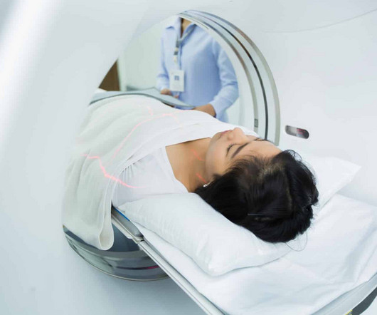
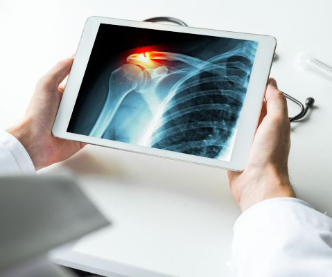
There is no need to be anxious as imaging tests are non-invasive and painless. For X-rays, it usually takes less than 10 minutes. The magneticresonanceimaging may take longer, depending on the area being scanned. X-ray Also called a radiograph, an X-ray uses radiation to create images of the body.
Effects of low-dose ionizing radiation on genomic instability in interventional radiology workers. Quantifying Regional Radiation-Induced Lung Injury in Patients Using Hyperpolarized 129Xe Gas Exchange MagneticResonanceImaging. Workplace violence in medical radiation science: A systematic review.
Food and Drug Administration (FDA) has said CT is still the preferred imaging modality for patients with medical devices. In an October 15 communication , the agency said it had received reports of electronic medical devices being damaged during CT scans due to radiation. Read the FDA's full guidance here.
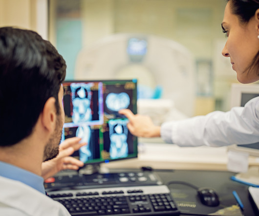
CT scans also aid in surgical planning as well as monitoring the effectiveness of cancer treatments like chemo or radiation therapy. Evaluating the Tissue and Organs in the Chest Chest CT scans are are more detailed than x-rays, giving you more information about possible diseases or injury of your chest organs.
Introduction: The history of X-rayimaging is a testament to the unceasing march of technology in healthcare. From the days of photographic film to the digital age, this blog traces the remarkable evolution of X-rayimaging, shedding light on how technology has transformed the practice of medicine.
Teleradiology Introduction: X-ray technology has been a cornerstone of modern medicine for over a century. This blog explores the evolution, significance, and the latest advancements in X-ray technology, shedding light on how it continues to shape and revolutionize the healthcare industry.
Initial X-Rays showed small osteophytes at the medial tibiotalar articulation with a small intra-articular body raising the possibility of anterior impingement. Postoperative X-Rays showed successful resection of anterior tibial and talar osteophytes with no further imaging evidence of impingement.
CT scans also aid in surgical planning as well as monitoring the effectiveness of cancer treatments like chemo or radiation therapy. By producing interior images of the coronary arteries, CT scans help identify blockages, aneurysms, or other abnormalities that can lead to damaging issues like a heart attack or stroke.
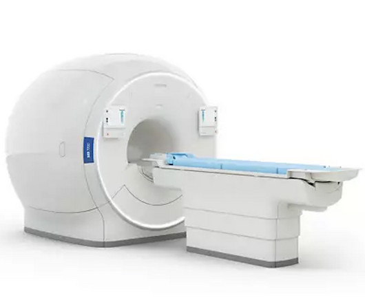
In this blog post, we’ll explore the differences and uses of MRI, CT scans, X-rays, ultrasounds, and PET/CT to help you better understand what to expect and how these technologies can assist in your healthcare. MagneticResonanceImaging (MRI) MRI stands for MagneticResonanceImaging.
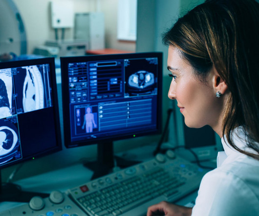
Medical imaging is a technology which is used by radiologists , particularly for diagnostic purposes. Although the word “radiology” sounds like it involves radiation, that is not always the case – for example, MRI (magneticresonanceimaging) and ultrasound do not use radiation in their medical imaging technologies.
As for magneticresonanceimaging (MRI), just 19% of extremely disadvantaged zip codes had access as compared to 32% of extremely advantaged. Low-dose X-ray Solutions Serve Broadest Patient Population Digital radiography has come a long way at Massac Memorial as well. Of these, rural zip codes totaled 1,160.
In 2011, a large study examined the use of x-rays and other radiationimaging on children—they estimated that the average child would get more than seven radiation scans by the age of 18. They analyze the images to identify abnormalities or signs of disease.
Medical imaging is used to help diagnose these injuries, so doctors can propose appropriate treatment plans. While X-rays are typically utilized, an MRI or CT scan may be recommended. To determine if the bone is broken, doctors may use one of the following imaging technologies. What Are Broken Bones?
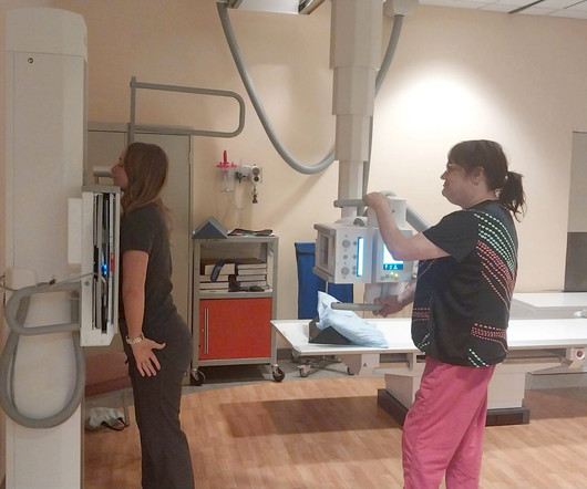
Each piece of technology encompassed in medical imaging focuses on a different area or system of the body. Take x-rays for example. X-rays are used to view the skeletal system – the bones – of the body. X-rays are used to identify different issues with a patient’s bones and joints.
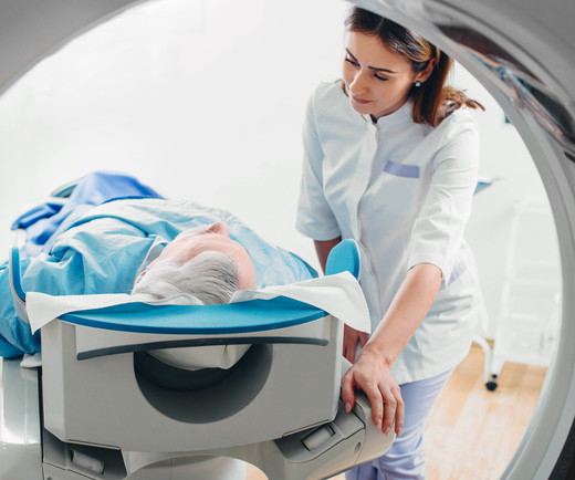
Magneticresonanceimaging (MRI) is used to help diagnose and treat various medical conditions. At Intermountain Medical Imaging , we rely on a variety of MRI options that offer a wider opening, helping us deliver high-quality care to clients and medical providers alike. MRIs rely on large magnets and radio waves.
Medical imaging is crucial in diagnosing and treating various medical conditions. These technologies have transformed the medical field from X-rays to MagneticResonanceImaging (MRI) and Computed Tomography ( CT ) scans.
Key Points: Cone Beam CT (CBCT) is superior in assessing bony structures compared to magneticresonanceimaging (MRI) In this study, there was a 40% rate of discrepancy when grading knee subchondral insufficiency fractures on CBCT vs. MRI, with MRI frequently underestimating damage of the subchondral bone plate while overestimating lesion size.
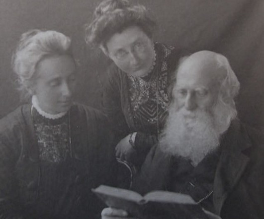
It all started when Wilhelm Conrad Röntgen discovered X-rays in 1895. After working for weeks in his lab experimenting on the production of ‘strange rays’, which he referred to as ‘X’, he asked his wife Anna Bertha to lend ‘a hand’, the left one to be precise, which he used to produce the first X-rayimage.
The Philips 3T MR 7700 and Polarean XENOVIEWTM (xenon Xe 129 hyperpolarized) enable an advanced solution for the evaluation of lung ventilation based on Xenon gas MR imaging, providing clinical confidence.
We organize all of the trending information in your field so you don't have to. Join 5,000 users and stay up to date on the latest articles your peers are reading.
You know about us, now we want to get to know you!
Let's personalize your content
Let's get even more personalized
We recognize your account from another site in our network, please click 'Send Email' below to continue with verifying your account and setting a password.


Let's personalize your content