This site uses cookies to improve your experience. To help us insure we adhere to various privacy regulations, please select your country/region of residence. If you do not select a country, we will assume you are from the United States. Select your Cookie Settings or view our Privacy Policy and Terms of Use.
Cookie Settings
Cookies and similar technologies are used on this website for proper function of the website, for tracking performance analytics and for marketing purposes. We and some of our third-party providers may use cookie data for various purposes. Please review the cookie settings below and choose your preference.
Used for the proper function of the website
Used for monitoring website traffic and interactions
Cookie Settings
Cookies and similar technologies are used on this website for proper function of the website, for tracking performance analytics and for marketing purposes. We and some of our third-party providers may use cookie data for various purposes. Please review the cookie settings below and choose your preference.
Strictly Necessary: Used for the proper function of the website
Performance/Analytics: Used for monitoring website traffic and interactions
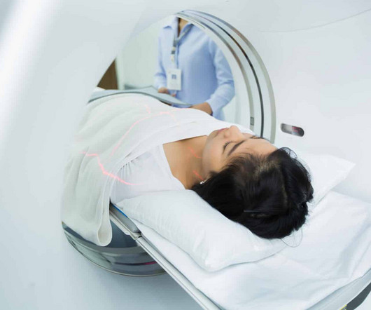
There is no need to be anxious as imaging tests are non-invasive and painless. For X-rays, it usually takes less than 10 minutes. The magneticresonanceimaging may take longer, depending on the area being scanned. X-ray Also called a radiograph, an X-ray uses radiation to create images of the body.
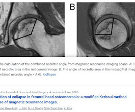
BACKGROUND The hypothesis is that the combined necrotic angle measurement from magneticresonanceimaging scans predicts the subsequent risk of collapse in hips with femoral head necrosis. With use of the modified method of Kerboul et al., With use of the modified method of Kerboul et al.,
Commercially Available Chest Radiograph AI Tools for Detecting Airspace Disease, Pneumothorax, and Pleural Effusion. Generative Artificial Intelligence for Chest Radiograph Interpretation in the Emergency Department. Cognitive Motor Dissociation in Disorders of Consciousness. To learn more about this paper, click here.
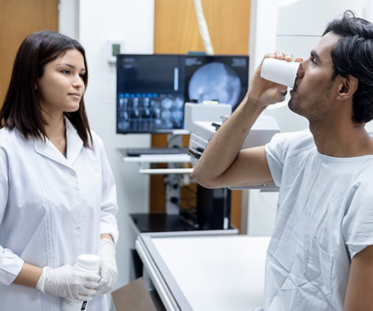
Injecting or drinking the media contrast helps doctors see blood vessels and organs more clearly in an x-ray or a computed tomography ( CT ) scan. Qureshi said these findings highlight the need to reduce the reliance on radiographic media contrast imaging without compromising patient outcomes.
The approval expands upon Bayer's focus on breast imaging, with a portfolio that also includes Gadavist (gadobutrol) injection, a gadolinium-based contrast agent approved for use with MRI ( MagneticResonanceImaging ) to assess the presence and extent of malignant breast disease in adult patients. for use in CEM.
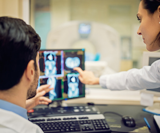
Medical imaging is a technology which is used by radiologists , particularly for diagnostic purposes. Although the word “radiology” sounds like it involves radiation, that is not always the case – for example, MRI (magneticresonanceimaging) and ultrasound do not use radiation in their medical imaging technologies.
It all started when Wilhelm Conrad Röntgen discovered X-rays in 1895. After working for weeks in his lab experimenting on the production of ‘strange rays’, which he referred to as ‘X’, he asked his wife Anna Bertha to lend ‘a hand’, the left one to be precise, which he used to produce the first X-rayimage.
Key Points: Imaging modalities such as plain radiographs (X-Ray), computed tomography (CT), and magneticresonanceimaging (MRI), dont have the diagnostic accuracy needed to detect syndesmotic widening or subtle instability.
We organize all of the trending information in your field so you don't have to. Join 5,000 users and stay up to date on the latest articles your peers are reading.
You know about us, now we want to get to know you!
Let's personalize your content
Let's get even more personalized
We recognize your account from another site in our network, please click 'Send Email' below to continue with verifying your account and setting a password.


Let's personalize your content