This site uses cookies to improve your experience. To help us insure we adhere to various privacy regulations, please select your country/region of residence. If you do not select a country, we will assume you are from the United States. Select your Cookie Settings or view our Privacy Policy and Terms of Use.
Cookie Settings
Cookies and similar technologies are used on this website for proper function of the website, for tracking performance analytics and for marketing purposes. We and some of our third-party providers may use cookie data for various purposes. Please review the cookie settings below and choose your preference.
Used for the proper function of the website
Used for monitoring website traffic and interactions
Cookie Settings
Cookies and similar technologies are used on this website for proper function of the website, for tracking performance analytics and for marketing purposes. We and some of our third-party providers may use cookie data for various purposes. Please review the cookie settings below and choose your preference.
Strictly Necessary: Used for the proper function of the website
Performance/Analytics: Used for monitoring website traffic and interactions
When reviewing radiographs, computed tomography (CT) scans or magneticresonanceimaging (MRI) scans, do you still turn to mnemonics every now and then to jog your short-term memory?
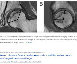
BACKGROUND The hypothesis is that the combined necrotic angle measurement from magneticresonanceimaging scans predicts the subsequent risk of collapse in hips with femoral head necrosis. With use of the modified method of Kerboul et al., With use of the modified method of Kerboul et al.,
Qureshi said these findings highlight the need to reduce the reliance on radiographic media contrast imaging without compromising patient outcomes. Some suggested methods include doing more magneticresonanceimaging ( MRI ) scans and reducing the amount of dye used per patient.
The approval expands upon Bayer's focus on breast imaging, with a portfolio that also includes Gadavist (gadobutrol) injection, a gadolinium-based contrast agent approved for use with MRI ( MagneticResonanceImaging ) to assess the presence and extent of malignant breast disease in adult patients. for use in CEM.
Medical imaging is a technology which is used by radiologists , particularly for diagnostic purposes. Although the word “radiology” sounds like it involves radiation, that is not always the case – for example, MRI (magneticresonanceimaging) and ultrasound do not use radiation in their medical imaging technologies.
Radiologists and radiographers undertaking the imaging have most likely never seen the patient before. The situation and the uncertainty about the coming results scared me but interaction with radiographers helped me through’: a qualitative study on patients’ experiences of magneticresonanceimaging examinations.
A) Dorsoplantar radiograph of the foot demonstrating an isolated fracture of the cuboid with possible extension into the tarsometatarsal joint. (B) B) Medial oblique radiograph of the foot demonstrating an isolated fracture of the cuboid. Radiographic evidence can support the diagnosis. Trauma in a 8 year old female.
A) AP radiograph of Lisfranc Fracture Dislocation demonstrates the circled “fleck sign” or Lisfranc ligament avulsion fracture fragment. (B) C) The lateral radiograph notes with a circle, the dorsal sub dislocation of the metatarsal base. Radiographs should be repeated after two weeks to ensure surgery is unnecessary.
Commercially Available Chest Radiograph AI Tools for Detecting Airspace Disease, Pneumothorax, and Pleural Effusion. Generative Artificial Intelligence for Chest Radiograph Interpretation in the Emergency Department. Cognitive Motor Dissociation in Disorders of Consciousness. To learn more about this paper, click here.
There is no need to be anxious as imaging tests are non-invasive and painless. The magneticresonanceimaging may take longer, depending on the area being scanned. Why Diagnostic Imaging Methods Are Important Medical imaging is used to diagnose, monitor, and treat medical problems.
after seeing the image. (2) Photoprint from radiograph by W.K. 3) In the early twentieth century, it was a common goal for investigators to try to find a way to separate the superimposed shadows that were recorded when a complex structure was shown on a radiograph. (3) This is now known as ‘Hand mit Ringen’. (1) Röntgen, 1895.
If this is where we are at already, it is not unreasonable to imagine that in 23 years AI will be capable of reporting imaging studies (CT or magneticresonanceimaging (MRI)) with a higher accuracy than humans and that these reports will no longer be routinely reported by human radiologists out of hours. Hough, Andrew.
It has well-defined radiographic features and various clinical presentations. They described six radiographic phenotypes of CSVD: (1) recent small subcortical infarct, (2) white matter hyperintensity, (3) lacune of presumed vascular origin, (4) widened perivascular spaces, (5) cerebral microbleed, and (6) brain atrophy.
Key Points: Imaging modalities such as plain radiographs (X-Ray), computed tomography (CT), and magneticresonanceimaging (MRI), dont have the diagnostic accuracy needed to detect syndesmotic widening or subtle instability.
Magneticresonanceimaging and computed tomography in emergency assessment of patients with suspected acute stroke: a prospective comparison. Radiographics. Approach to reperfusion therapy for acute ischemic stroke. Last Update: September 14, 2018. Accessed from [link] Chaela JA, Kidwell CS, Nentwich LM, et al.
We organize all of the trending information in your field so you don't have to. Join 5,000 users and stay up to date on the latest articles your peers are reading.
You know about us, now we want to get to know you!
Let's personalize your content
Let's get even more personalized
We recognize your account from another site in our network, please click 'Send Email' below to continue with verifying your account and setting a password.


Let's personalize your content