This site uses cookies to improve your experience. To help us insure we adhere to various privacy regulations, please select your country/region of residence. If you do not select a country, we will assume you are from the United States. Select your Cookie Settings or view our Privacy Policy and Terms of Use.
Cookie Settings
Cookies and similar technologies are used on this website for proper function of the website, for tracking performance analytics and for marketing purposes. We and some of our third-party providers may use cookie data for various purposes. Please review the cookie settings below and choose your preference.
Used for the proper function of the website
Used for monitoring website traffic and interactions
Cookie Settings
Cookies and similar technologies are used on this website for proper function of the website, for tracking performance analytics and for marketing purposes. We and some of our third-party providers may use cookie data for various purposes. Please review the cookie settings below and choose your preference.
Strictly Necessary: Used for the proper function of the website
Performance/Analytics: Used for monitoring website traffic and interactions
They do not include any physician work value, but they would be available in the imaging center using global billing. 55881 Ablation of prostate tissue, transurethral, using thermal ultrasound, including magneticresonanceimaging guidance for, and monitoring of, tissue ablation. PC-6.47 $525.63 $209.28
The work included data from eight Google searches related to a medical imaging modality such as x-rays, CT scans, MRIs, PET scans, CT and MR angiography, and ultrasounds. A search for "Radiology Chest X-ray filetype:ppt" resulted in accessible PHI in 40% of the presentations with images. contained full PHI.
Given the increasing prevalence of breast reconstruction, researchers discussed key signs of common complications on magneticresonanceimaging (MRI), ultrasound and other imaging during a recent presentation at the American Roentgen Ray Society (ARRS) 2023 Annual Meeting in Honolulu, Hawaii.
Professional Radiology strives to provide patients with accurate and compassionate imaging services. If you’re looking for diagnostic imaging services in the El Paso region, contact us online or call (915) 225-2480 to schedule an appointment today. It is simple and quick to use, often taking less than 30 minutes to complete.
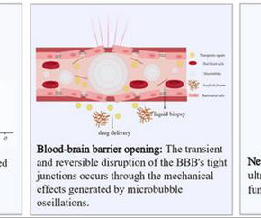
milla1cf Mon, 04/08/2024 - 20:52 April 8, 2024 — Magneticresonance-guided focused ultrasound (MRgFUS) is a non-invasive technique for neuroregulation. Currently, the U.S.
A new study using machine learning to automatically digitally combine the diagnostic capabilities of pelvic scans and magneticresonanceimaging (MRI) for identifying endometriosis lesions seeks to shorten the diagnostic journey as well as reduce reliance on surgery.
A new study using machine learning to automatically digitally combine the diagnostic capabilities of pelvic scans and magneticresonanceimaging (MRI) for identifying endometriosis lesions seeks to shorten the diagnostic journey as well as reduce reliance on surgery.
Imaging techniques such as computed tomography, magneticresonanceimaging, positron emission tomography and ultrasound have become indispensable in the medical world.
Offer Versatility and Clarity to Your Doctors and Patients MRI, or MagneticResonanceImaging , could be the most versatile imaging technology available to members of the medical community. The answer to many of these questions could be just one—MRI.
The innovative technology aims to be more accurate as well as cheaper to provide than today's most common diagnostic tools such as X-ray mammography, ultrasound and magneticresonanceimaging (MRI).
Offering the full modality of diagnostic scans, including 3T MRI, CT, ultrasound, 3-D mammography, PET/CT, nuclear medicine, DEXA, X-rays, among others, the company uses the newest, most advanced, diagnostic imaging technologies while maintaining affordability and accessibility.
However, concerns about high perioperative blood loss and long-term side effects of ejaculatory dysfunction, impotency, and urethral strictures have driven the innovation of numerous minimally invasive alternatives, including prostatic artery embolization (PAE), transurethral ultrasound ablation, transurethral water vapor therapy, water-jet ablation, (..)
Helps Doctors Monitor Disease Progression Diagnostic imaging is one of the most important markers that doctors utilize to monitor the progression of diseases for a variety of ailments, including: Certain types of malignant cancer Pneumonia and other respiratory diseases Internal bleeding Brain injuries Cardiac conditions Acute injuries (i.e.,
It offers a product and solution portfolio for all key diagnostic imaging modalities: X-ray imaging (including Computed Tomography-CT, Interventional Radiology, and Cardiac Catheterization), MagneticResonanceImaging (MRI), Contrast Enhanced Ultrasound (CEUS), and Nuclear Medicine through radioactive tracers and novel PET imaging agents to inform (..)
During the diagnostic process, medical imaging assists with assessing the blood vessels and surrounding tissue. Learn how CT scans and magneticresonanceimaging (MRI) play a role. A transcranial Doppler ultrasound can better illustrate blood circulation within the brain. What Is a Stroke?
PSMA-Targeted Nanobubbles (PSMA-NB) are a promising clinical tool for increasing the accuracy of prostate biopsy in situations where multiparametric magneticresonanceimaging (mpMRI) is not available.
Whether it’s through X-ray, MRI, CT scan, ultrasound, or using tiny cameras, health care professionals can see beyond the flesh and gain a deeper understanding of the human body’s internal machinations. These imaging methods diagnose all kinds of conditions, but can often be intimidating.
Urology: GE HealthCare’s ultrasound guidance technology combined with MIM Software’s post-processing and fusion expertise yield’s an integrated suite of prostate fusion solutions with the ability to address needs across the care pathway to support a patient's personal prostate cancer journey. Ask your local representative about your options.
Which of the following imaging modalities is indicated at this time? Barium contrast X-ray CT scan MagneticresonanceimagingUltrasound FOR THE RIGHT ANSWER CLICK ON THE ROSH REVIEW LOGO BELOW References Burkart JM, Bleyer A. Peritoneal catheter exit-site and tunnel infections in peritoneal dialysis in adults.
Conventional B-mode ultrasound is not sensitive to many cancers, including prostate. The incorporation of magneticresonanceimaging (MRI) to detect areas of suspicion for prostate cancer along with hardware-based MRI-ultrasoundimage fusion platforms allows physicians to biopsy MRI targets in an office setting, outside of an MRI suite.
Medical imaging is a technology which is used by radiologists , particularly for diagnostic purposes. Although the word “radiology” sounds like it involves radiation, that is not always the case – for example, MRI (magneticresonanceimaging) and ultrasound do not use radiation in their medical imaging technologies.
Magneticresonanceimaging-guided high intensity focused ultrasound (MR-HIFU) allows precise, image-guided and completely non-invasive ablation without ionizing radiation and has been shown by several groups, including ours, to be a good treatment alternative to RFA.
The imaging evaluation of chronic mesenteric ischemia (CMI) is usually first performed with a duplex ultrasound study. As seen with other disease patterns, many operators subsequently obtain cross-sectional or dynamic imaging for further evaluation.
In this blog post, we’ll explore the differences and uses of MRI, CT scans, X-rays, ultrasounds, and PET/CT to help you better understand what to expect and how these technologies can assist in your healthcare. MagneticResonanceImaging (MRI) MRI stands for MagneticResonanceImaging.
The product can be used to visualize known or suspected lesions of the breast in adults, as an adjunct to mammography and/or ultrasound. 1 The new FDA approved indication aligns with the recent increased focus on supplemental imaging needs for women at a higher risk for breast cancer , which may include the 40-50% of U.S.
Breast Ultrasound This screening is not usually performed as the routine test; it is often a follow-up to a mammogram in which an abnormality was detected. For example, women with breast lumps may have an ultrasound to determine more detailed characteristics of the abnormality. Ultrasound works by emitting sound waves through the skin.
Section 2: Extraneous Imaging Defined: Define extraneous imaging in the context of trauma radiology, exploring the various imaging modalities beyond conventional radiography and computed tomography (CT) scans.
DIS is a full-service independent imaging center that performs both standard and advanced screening and diagnostic scans. Click here to view the Edge of the Lake Magazine issue that lists the top winners – including Diagnostic Imaging Services. Six locations serve the contiguous tri-parish areas of Orleans, Jefferson and St.
Blood tests Imaging tests Physical exams What testing can be used to diagnose thyroid disorders? Computerized Tomography (CT) UltrasoundMagneticResonanceImaging (MRI) Biopsy Nuclear scan/radioactive iodine uptake What are some signs that you need to speak to your doctor about your thyroid?
Technological Metamorphosis: From Analog to Digital Prowess Chart the transition from analog radiology to the epoch of digital imaging technologies. Spotlight key technological breakthroughs, including computed tomography (CT), magneticresonanceimaging (MRI), and ultrasound, revolutionizing the diagnostic panorama.
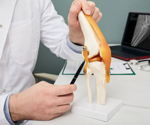
For diagnostic purposes, your doctor may recommend undergoing magneticresonanceimaging (MRI). If clear differences are present, your doctor might make a definitive diagnosis or schedule imaging to confirm an ACL injury. Yet this condition can also affect amateur athletes and weekend warriors. What Is an ACL Injury?
Other types of medical imaging includes ultrasounds, MagneticResonanceImaging (MRI), Computed Tomography (CT), and even mammography. Medical imaging, as a practice, has been around since the last 19th century. Muyinatu Bell Muyinatu Bell is a modern counterpart of the forerunners of medical imaging.
The dye infiltrates internal tissue, so that images from the CT scan are more pronounced and clearer. An MRI or magneticresonanceimaging produces detailed images of the kidneys using radio waves. Doctors are able to diagnose and stage the progression of kidney cancer using the images produced by an MRI.
MRIs, also known as MagneticResonanceImaging, allow for very detailed scans with the help of a magnetic field and pulses of radio wave energy. They help doctors see organs and structures inside the body that is different than other types of scans such as X-rays, ultrasounds, or CT scans. What is a 3T MRI?
Depending on the diagnosis and severity of the patient’s condition, a physician may use a wide array of imaging tests to help determine the best cancer treatment. This method of imaging is most frequently used to look for cancer in the abdomen as well as guiding a biopsy in breast tissue.
The role of a pediatric radiologist involves the following: Interpreting Imaging Studies: Pediatric radiologists review and interpret various imaging studies such as X-rays, ultrasounds, computed tomography (CT) scans, magneticresonanceimaging (MRI), and nuclear medicine scans.
Types of Imaging Studies That Often Need a Second Opinion Not all imaging studies are straightforward, and some require deeper insight to reach a definitive diagnosis. Our radiologists with breast imaging expertise can re-evaluate mammograms, breast MRIs, and ultrasounds.
Some women do not realize they have them unless they are spotted in a routine exam or ultrasound. These tests include: Ultrasound Lab Tests MagneticResonanceImaging (MRI) Hysterosonography Hysterosalpingography Hysteroscopy If you experience any symptoms or have a family history of uterine fibroids, speak to your doctor.
Tests we can use in diagnosing your condition and monitoring the healing process include: Computerized tomography (CT) scanning, which provides cross-dimensional images of various body parts; Magneticresonanceimaging (MRI), which rely on magnets and radio waves to create moving images of various body parts and processes; Ultrasound scans, which use (..)
For an abdominal ultrasound: Hospitals charge $461 plus additional fees. For magneticresonanceimaging (MRI): Depending on whether contrast dye is used, hospitals charge anywhere between $1,800 and $3,000 plus other costs. At IMI, you pay between $650 and $800. At IMI, your total price is $379.
Delving Deeper into CPACS The heart of CPACS lies in its ability to collect, manage, and store all cardiology-related images. These include images from various modalities such as echocardiography, nuclear cardiology, vascular ultrasound, electrocardiography (ECG), and others.
This standard has revolutionized the radiology industry, encompassing many imaging modalities such as X-rays, computed tomography (CT), magneticresonanceimaging (MRI), ultrasound, nuclear medicine, PET scans, etc.
Image from Nico Sollmann, MD, PhD, of University Hospital Ulm and University Hospital Rechts der Isar, et al. Image from Shilpa Vijayakumar, MD, of Brigham and Women’s Hospital, et al. Ultrasound model predicts liver disease progression. Image from Fangyi Liu, MD, of the Chinese PLA General Hospital, et al.
We organize all of the trending information in your field so you don't have to. Join 5,000 users and stay up to date on the latest articles your peers are reading.
You know about us, now we want to get to know you!
Let's personalize your content
Let's get even more personalized
We recognize your account from another site in our network, please click 'Send Email' below to continue with verifying your account and setting a password.


Let's personalize your content