This site uses cookies to improve your experience. To help us insure we adhere to various privacy regulations, please select your country/region of residence. If you do not select a country, we will assume you are from the United States. Select your Cookie Settings or view our Privacy Policy and Terms of Use.
Cookie Settings
Cookies and similar technologies are used on this website for proper function of the website, for tracking performance analytics and for marketing purposes. We and some of our third-party providers may use cookie data for various purposes. Please review the cookie settings below and choose your preference.
Used for the proper function of the website
Used for monitoring website traffic and interactions
Cookie Settings
Cookies and similar technologies are used on this website for proper function of the website, for tracking performance analytics and for marketing purposes. We and some of our third-party providers may use cookie data for various purposes. Please review the cookie settings below and choose your preference.
Strictly Necessary: Used for the proper function of the website
Performance/Analytics: Used for monitoring website traffic and interactions
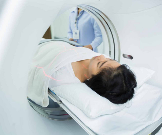
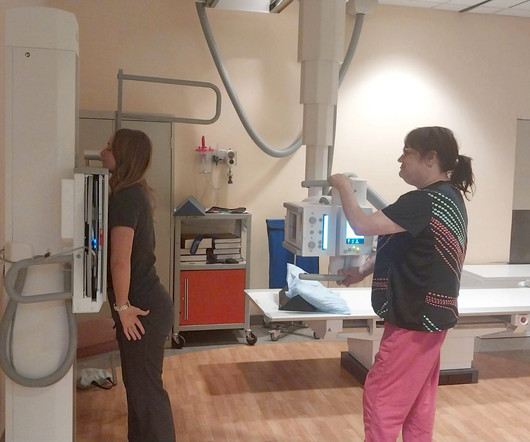
There is no need to be anxious as imaging tests are non-invasive and painless. For X-rays, it usually takes less than 10 minutes. The magneticresonanceimaging may take longer, depending on the area being scanned. X-ray Also called a radiograph, an X-ray uses radiation to create images of the body.
The work included data from eight Google searches related to a medical imaging modality such as x-rays, CT scans, MRIs, PET scans, CT and MR angiography, and ultrasounds. A search for "Radiology Chest X-ray filetype:ppt" resulted in accessible PHI in 40% of the presentations with images. contained full PHI.
Professional Radiology strives to provide patients with accurate and compassionate imaging services. If you’re looking for diagnostic imaging services in the El Paso region, contact us online or call (915) 225-2480 to schedule an appointment today.
On November 29, the company will present safety data for Elucirem in an abstract titled "Safety of gadopiclenol for magneticresonanceimaging (MRI): a pooled analysis of eight studies," it said.
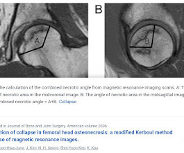
BACKGROUND The hypothesis is that the combined necrotic angle measurement from magneticresonanceimaging scans predicts the subsequent risk of collapse in hips with femoral head necrosis. With use of the modified method of Kerboul et al., With use of the modified method of Kerboul et al.,
However, "for imaging procedures specifically, CT continues to be the preferred tomographic imaging technology for people with implantable or wearable medical devices. CT is safer than magneticresonanceimaging (MRI) for people with devices of unknown MRI safety status." Read the FDA's full guidance here.
Quantifying Regional Radiation-Induced Lung Injury in Patients Using Hyperpolarized 129Xe Gas Exchange MagneticResonanceImaging. breast cancer detection software, Hologic Grace multimodal foundation model for medical imaging, HOPPR HealthFLD liver attenuation AI software, Nanox.ai
Evaluating the Tissue and Organs in the Chest Chest CT scans are are more detailed than x-rays, giving you more information about possible diseases or injury of your chest organs. CT scans also aid in surgical planning as well as monitoring the effectiveness of cancer treatments like chemo or radiation therapy.
The innovative technology aims to be more accurate as well as cheaper to provide than today's most common diagnostic tools such as X-ray mammography, ultrasound and magneticresonanceimaging (MRI).
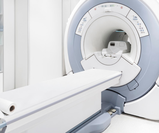
Offer Versatility and Clarity to Your Doctors and Patients MRI, or MagneticResonanceImaging , could be the most versatile imaging technology available to members of the medical community. The answer to many of these questions could be just one—MRI.
Introduction: The history of X-rayimaging is a testament to the unceasing march of technology in healthcare. From the days of photographic film to the digital age, this blog traces the remarkable evolution of X-rayimaging, shedding light on how technology has transformed the practice of medicine.
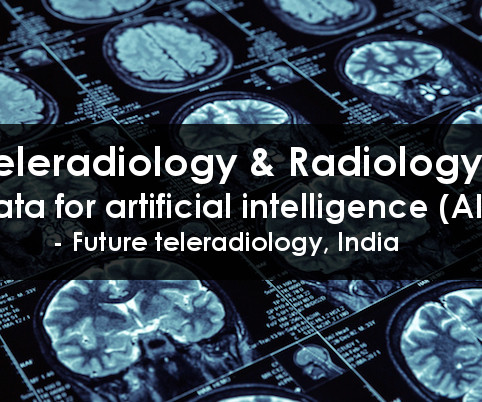
Teleradiology Introduction: X-ray technology has been a cornerstone of modern medicine for over a century. This blog explores the evolution, significance, and the latest advancements in X-ray technology, shedding light on how it continues to shape and revolutionize the healthcare industry.
milla1cf Fri, 11/03/2023 - 12:22 November 3, 2023 — Guerbet , a global leader in medical imaging with more than 30 years of experience in MRI, announced today its schedule of activities at the 2023 Radiological Society of North America (RSNA) Scientific Assembly and Annual Meeting from November 26-30 in Chicago, IL.
Initial X-Rays showed small osteophytes at the medial tibiotalar articulation with a small intra-articular body raising the possibility of anterior impingement. Postoperative X-Rays showed successful resection of anterior tibial and talar osteophytes with no further imaging evidence of impingement.
milla1cf Thu, 01/04/2024 - 10:47 January 4, 2024 — Diagnosing cancer today involves using chemical “contrast agents” to improve the accuracy of medical imaging processes such as X-rays as well as computed tomography (CT) and magneticresonanceimaging (MRI) scans.
Helps Doctors Monitor Disease Progression Diagnostic imaging is one of the most important markers that doctors utilize to monitor the progression of diseases for a variety of ailments, including: Certain types of malignant cancer Pneumonia and other respiratory diseases Internal bleeding Brain injuries Cardiac conditions Acute injuries (i.e.,
Bracco Imaging S.p.A., part of the Bracco Group, is a global diagnostic imaging provider, headquartered in Milan, Italy, which develops, manufactures and markets diagnostic imaging agents and solutions.
Injecting or drinking the media contrast helps doctors see blood vessels and organs more clearly in an x-ray or a computed tomography ( CT ) scan. Qureshi said these findings highlight the need to reduce the reliance on radiographic media contrast imaging without compromising patient outcomes.
In this blog post, we’ll explore the differences and uses of MRI, CT scans, X-rays, ultrasounds, and PET/CT to help you better understand what to expect and how these technologies can assist in your healthcare. MagneticResonanceImaging (MRI) MRI stands for MagneticResonanceImaging.
Which of the following imaging modalities is indicated at this time? Barium contrast X-ray CT scan Magneticresonanceimaging Ultrasound FOR THE RIGHT ANSWER CLICK ON THE ROSH REVIEW LOGO BELOW References Burkart JM, Bleyer A. McGraw Hill; 2020:(Ch) 90. link] Szeto CC, Li PKT. Clin J Am Soc Nephrol.
Whether it’s through X-ray, MRI, CT scan, ultrasound, or using tiny cameras, health care professionals can see beyond the flesh and gain a deeper understanding of the human body’s internal machinations.
As for magneticresonanceimaging (MRI), just 19% of extremely disadvantaged zip codes had access as compared to 32% of extremely advantaged. Low-dose X-ray Solutions Serve Broadest Patient Population Digital radiography has come a long way at Massac Memorial as well. Of these, rural zip codes totaled 1,160.
MRI Isn’t An Option Another form of diagnostic imaging is MagneticResonanceImaging (MRI) , which is used to diagnose brain and spinal cord disorders, joint and musculoskeletal conditions, and cancer. In the case of an MRI, this method may not be used on patients who have metallic objects in their bodies.
Gadolinium alginate hydrogel consists of biocompatible alginate polymers cross-linked by gadolinium ions which confer magneticresonanceimaging (MRI) and X-ray contrast enhancement. To develop gadolinium alginate-based hydrogel as embolic agent and explore its applications in theranostics.
Medical imaging is a technology which is used by radiologists , particularly for diagnostic purposes. Although the word “radiology” sounds like it involves radiation, that is not always the case – for example, MRI (magneticresonanceimaging) and ultrasound do not use radiation in their medical imaging technologies.
Medical imaging is used to help diagnose these injuries, so doctors can propose appropriate treatment plans. While X-rays are typically utilized, an MRI or CT scan may be recommended. To determine if the bone is broken, doctors may use one of the following imaging technologies. What Are Broken Bones?
The approval expands upon Bayer's focus on breast imaging, with a portfolio that also includes Gadavist (gadobutrol) injection, a gadolinium-based contrast agent approved for use with MRI ( MagneticResonanceImaging ) to assess the presence and extent of malignant breast disease in adult patients. for use in CEM.
In particular, AI/ML models are finding increasing applications in the analysis of medical images. This includes X-ray , computed tomography , and magneticresonanceimages.
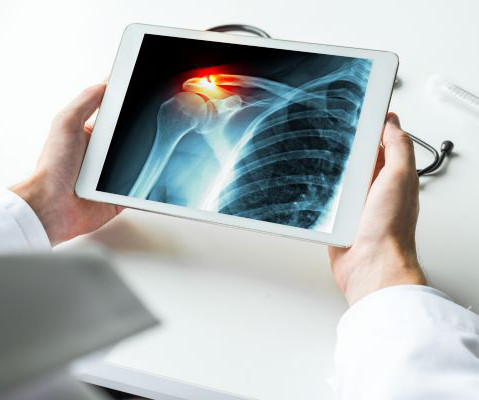
Each piece of technology encompassed in medical imaging focuses on a different area or system of the body. Take x-rays for example. X-rays are used to view the skeletal system – the bones – of the body. X-rays are used to identify different issues with a patient’s bones and joints.
Magneticresonanceimaging (MRI) is used to help diagnose and treat various medical conditions. At Intermountain Medical Imaging , we rely on a variety of MRI options that offer a wider opening, helping us deliver high-quality care to clients and medical providers alike. MRIs rely on large magnets and radio waves.
There are several types of imaging tests that physicians use to detect cancer in patients: X-Ray, Computed Tomography (CT), MagneticResonanceImaging (MRI), Ultrasound (US), Nuclear Medicine, and Positron Emission Tomography (PET). It is also used to determine the progression of existing cancerous masses.
DIS is a full-service independent imaging center that performs both standard and advanced screening and diagnostic scans. Capitol Imaging Services is the largest independent radiology practice of its type serving the Southeastern United States. Six locations serve the contiguous tri-parish areas of Orleans, Jefferson and St.
In 2011, a large study examined the use of x-rays and other radiation imaging on children—they estimated that the average child would get more than seven radiation scans by the age of 18. They analyze the images to identify abnormalities or signs of disease.
Medical imaging is crucial in diagnosing and treating various medical conditions. These technologies have transformed the medical field from X-rays to MagneticResonanceImaging (MRI) and Computed Tomography ( CT ) scans.
Inception and Exploration: Revealing the Genesis Explore the formative days of radiology, ignited by Wilhelm Roentgen’s groundbreaking discovery of X-rays in 1895. Technological Metamorphosis: From Analog to Digital Prowess Chart the transition from analog radiology to the epoch of digital imaging technologies.
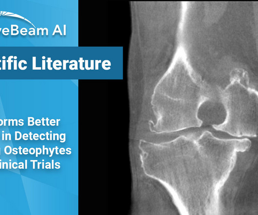
Key Points: Cone Beam CT (CBCT) is superior in assessing bony structures compared to magneticresonanceimaging (MRI) In this study, there was a 40% rate of discrepancy when grading knee subchondral insufficiency fractures on CBCT vs. MRI, with MRI frequently underestimating damage of the subchondral bone plate while overestimating lesion size.
For diagnostic purposes, your doctor may recommend undergoing magneticresonanceimaging (MRI). If clear differences are present, your doctor might make a definitive diagnosis or schedule imaging to confirm an ACL injury. Yet this condition can also affect amateur athletes and weekend warriors. What Is an ACL Injury?
Key Points: While magneticresonanceimaging (MRI) adequately detects the size and presence of osteophytes (OPs) in the medial tibio-femoral compartment, it underestimates OPs in all other knee compartments of osteoarthritic patients. In the research setting, weight bearing CT (WBCT) imaging could be a useful tool.
Closeup of X-ray photography of human brain Introduction: MagneticResonanceImaging (MRI) professionals are at the heart of the medical imaging world, shaping the diagnosis and care of countless patients.
MRIs, also known as MagneticResonanceImaging, allow for very detailed scans with the help of a magnetic field and pulses of radio wave energy. They help doctors see organs and structures inside the body that is different than other types of scans such as X-rays, ultrasounds, or CT scans. What is a 3T MRI?
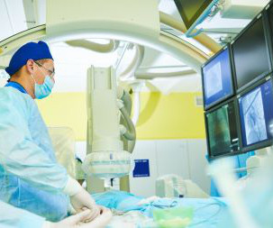
Subspecialized Expertise Consider the wide-ranging scope of your medical imaging needs. At various times, you may need to rely on an expert to interpret images produced by X-ray, magneticresonanceimaging (MRI), computed tomography (CT), positron emission tomography/computed tomography (PET/CT), or another modality.
It all started when Wilhelm Conrad Röntgen discovered X-rays in 1895. After working for weeks in his lab experimenting on the production of ‘strange rays’, which he referred to as ‘X’, he asked his wife Anna Bertha to lend ‘a hand’, the left one to be precise, which he used to produce the first X-rayimage.
Tests we can use in diagnosing your condition and monitoring the healing process include: Computerized tomography (CT) scanning, which provides cross-dimensional images of various body parts; Magneticresonanceimaging (MRI), which rely on magnets and radio waves to create moving images of various body parts and processes; Ultrasound scans, which use (..)
We invite you to visit our page detailing hospital imaging costs versus IMI’s charges. The following are some examples of how our fees compare to those of local hospitals*: For a two-view chest x-ray: Hospitals charge a minimum of $198. At IMI, you pay $70. At IMI, you pay between $650 and $800.
We organize all of the trending information in your field so you don't have to. Join 5,000 users and stay up to date on the latest articles your peers are reading.
You know about us, now we want to get to know you!
Let's personalize your content
Let's get even more personalized
We recognize your account from another site in our network, please click 'Send Email' below to continue with verifying your account and setting a password.


Let's personalize your content