This site uses cookies to improve your experience. To help us insure we adhere to various privacy regulations, please select your country/region of residence. If you do not select a country, we will assume you are from the United States. Select your Cookie Settings or view our Privacy Policy and Terms of Use.
Cookie Settings
Cookies and similar technologies are used on this website for proper function of the website, for tracking performance analytics and for marketing purposes. We and some of our third-party providers may use cookie data for various purposes. Please review the cookie settings below and choose your preference.
Used for the proper function of the website
Used for monitoring website traffic and interactions
Cookie Settings
Cookies and similar technologies are used on this website for proper function of the website, for tracking performance analytics and for marketing purposes. We and some of our third-party providers may use cookie data for various purposes. Please review the cookie settings below and choose your preference.
Strictly Necessary: Used for the proper function of the website
Performance/Analytics: Used for monitoring website traffic and interactions
A radiologist’s perception when viewing a complex MR image may be akin to a Major League Baseball (MLB) batter reading the stitches on a fastball, according to researchers exploring exactly how diagnostic interpretations are made. The Perception Lab is a "pop-up" version of an academic research lab focused on medicalimage perception.
While the pandemic affected medical operations across the country, the experts said that radiologists developed and honed their sense of resiliency as imaging was placed on the front lines. Medicalimaging played a significant role in the early days of the pandemic when it hit its initial peak in April 2020.
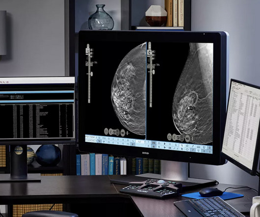
Healthcare disparities continue to plague medicalimaging, but there are concrete measures radiologists can take to mitigate them, according to a paper published on October 12 in RadioGraphics. B) A craniocaudal magnified mammogram more clearly shows the irregular mass and pleomorphic calcifications in the medial breast. (C)
The work presented at RSNA builds on previous research that investigated the feasibility of adversarial attacking (black-box type), in which data generated by generative adversarial networks (GANs) are inserted into the image as either positive- or negative-looking adversarial image features.
Everything we do at DeepHealth is about empowering radiologists and healthcare professionals, not just in the detection and diagnosis of diseases but across the whole care continuum. Saige Breast has been trained on over 10,000 scans and has now been employed in over a million mammograms. What is DeepHealth doing this year at ECR?
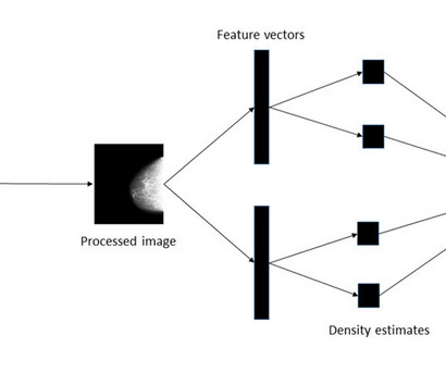
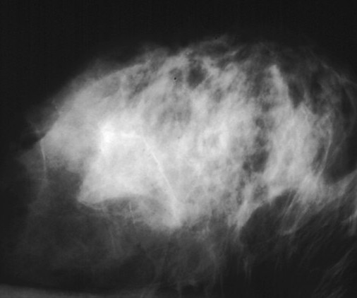
Why Breast Density Matters in Cancer Screening Dense breast tissue affects screening in two key ways: Reduced Visibility : Dense tissue appears white on mammograms, as do tumors, making it harder to detect abnormalities. Inter-radiologist Variation : Assessments can vary up to 33% 1 when different radiologists interpret the same mammograms.
With AI-mediated cyberattacks on the rise, radiologists can find it challenging to balance prompt patient access to diagnostic imaging while safeguarding sensitive healthcare data. With growing radiology practices and outpatient imaging facilities, we will see an increase in outsourced diagnostic imaging. Dhaval Shah.
An artificial intelligence (AI) system, which mimics the gaze of radiologists reading medicalimages such as mammograms, has been developed by a team of scientists at Cardiff University.
A new AI system that mimics the gaze of radiologists interpreting medicalimages, such as mammograms, can enhance the speed, precision, and sensitivity of medical diagnostics while facilitating early detection of breast cancer.
A new AI system that mimics the gaze of radiologists interpreting medicalimages, such as mammograms, can enhance the speed, precision, and sensitivity of medical diagnostics while facilitating early detection of breast cancer.
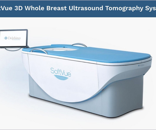
mtaschetta-millane Tue, 07/02/2024 - 09:50 July 2, 2024 — Delphinus Medical Technologies , a pioneering medicalimaging company that developed the SoftVue Breast Ultrasound Tomography (UST), announced today the publication of a study comparing mammography in conjunction with SoftVue UST vs mammography alone in women with dense breasts.
MedicalImaging of Fredericksburg Ranked First Place in “Best of the Burg” 2019 Contest MedicalImaging of Fredericksburg (MIF) took first place in the 2019 “Best of the Burg” contest, earning the award for “Best MedicalImaging”.
While various methods are available to estimate this measure, studies have shown that subjective assessments conducted by radiologists based on visual analogue scales are more accurate than any other method. Their findings are published in the Journal of MedicalImaging. “The
As we are turning the corner in the Fredericksburg region from COVID-19, MedicalImaging of Fredericksburg is pleased to resume scheduled appointments at all six of our locations. On May 4th, MedicalImaging of Fredericksburg will reopen all of its facilities for scheduled appointments.
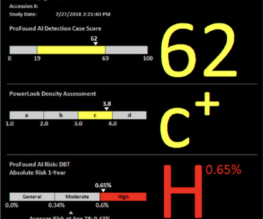
The company will also highlight its recently launched ProFound Cloud, an advanced software-as-a-service (SaaS) platform that offers medical providers a cost-effective, secure, and scalable solution for accessing and deploying the latest ProFound Breast Health Suite of AI solutions. Mark Traill , M.D., unit increase in the score.
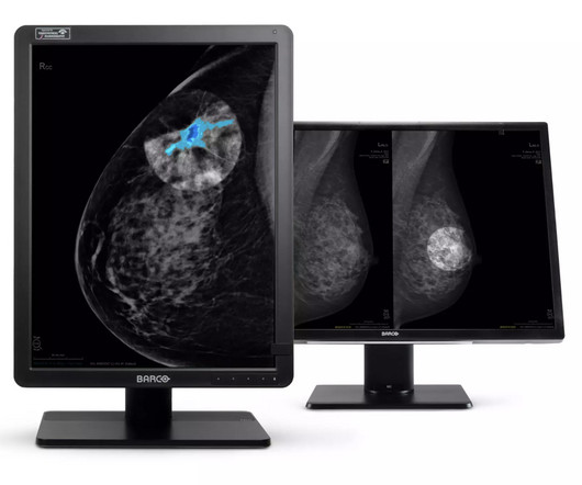
1 LG Business Solutions has set out to reduce the risk of this hardship with advanced medical monitor solutions. With the latest high-end medical display technology from LG, physicians can analyze radiological and mammogramimages with complete confidence. That leaves 9.5%
However, this can make it a bit trickier to detect breast cancer with a standard mammogram. Your breast density is something your radiologist determines when they read your mammogram. This information is included in your mammogram results, so be sure to ask your doctor about your breast density.
In the United States alone, it is estimated that around 40 million mammograms were performed each year. Mammograms are crucial as they are the primary method for early detection of breast cancer, enabling timely intervention and improving survival rates.
Try as we might, good health practices tend to be neglected, especially regarding preventive health screenings like mammograms. Research shows that one in four women who should be getting regular mammograms don’t. Women with an average risk for breast cancer should begin mammogram screening every year starting at 40.
Two breast radiologists reviewed PEM images taken one and four hours post 18 F-FDG injection and correlated the findings with lab results. For the study, 25 women, median age 52, recently diagnosed with breast cancer, underwent low-dose PEM with the radiotracer fluorine 18-labeled fluorodeoxyglucose ( 18 F-FDG).
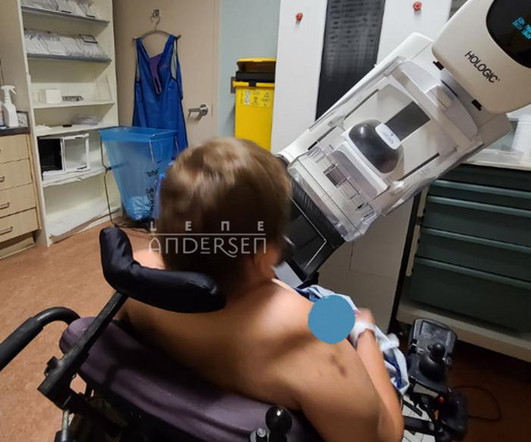
Her story is featured in an upcoming themed issue of the Journal of MedicalImaging and Radiation Sciences on the topic of specialized populations, published by Elsevier. Despite having a lump in her breast for several decades, she was unable to undergo mammograms due to the lack of accessible equipment and procedures.
“The existing Intuitive Workflow Tools will benefit from this add- on AI software tool which addresses a significant unmet need in medicalimaging, enabling rapid and accurate lesion segmentation, measurement, and visualization” says Dirk Feyants , Executive Vice President of Diagnostic Imaging at Barco.
The pioneer behind 3D mammography , Hologic will present new data that showcase how next-generation deep-learning solutions can assist with breast cancer detection and improve workflow for radiologists. Presented by esteemed breast imaging specialist and study author Dr. Sarah M.
This often involves blood, urine, and DNS tests, and medicalimaging studies like mammograms, CT lung cancer screening scans and CT virtual colonography. At Capitol Imaging Services, a board-certified radiologist is assigned to each imaging test for cancer we perform throughout our network.
An AI model developed by Duke University researchers can predict five-year breast cancer risk from mammograms, a study published March 19 in Radiology found. We hope that this [model] will enable some of those personalized screening strategies to reduce the load on patients and radiologists,” Donnelly told AuntMinnie.com. “We
Just as screening mammography continues to be the gold standard in terms of breast cancer detection, ACR Accreditation is also considered the gold standard in ensuring that patients receive the highest level of image quality and safety. This also means that we may put the deficiency or failure on our shoulders, as well.
Since then over 700 algorithms in medicalimaging have received U.S. There are several barriers that still need to be addressed from a radiologist, facility, and patient standpoint. If the cost is low (less than $10), the facility or radiologist absorbing this, while not optimal, makes it pretty much a non-issue.
Advanced technology converges with local history as MedicalImaging of Fredericksburg partners with Washington Heritage Museums in spreading breast cancer awareness this October. Ribbons are available at Mary Washington House and any of the five MedicalImaging of Fredericksburg locations.
That decline has been attributed, in large part, to annual screening mammograms. Each year, during your mammogram appointment, a full assessment is done to determine if you have one or multiple risk factors. A mammogram can show a mass before you can even feel it. The #1 reason why your annual mammogram can save your life.
On behalf of ACR — representing more than 41,000 diagnostic radiologists, interventional radiologists, radiation oncologists, nuclear medicine physicians and medical physicists, he urged the Subcommittee to provide at least $50.9 billion to the NIH for fiscal year (FY) 2024, an increase of $3.5
IR came to life through the combination of the creative thinking and technical skills of diagnostic radiologists and angiogiographers. (11) 11) Charles Dotter, a vascular radiologist and commonly known as ‘The Father of Interventional Radiology’, invented angioplasty. (12) 15) Radiologists needed a common means for sharing images.
We organize all of the trending information in your field so you don't have to. Join 5,000 users and stay up to date on the latest articles your peers are reading.
You know about us, now we want to get to know you!
Let's personalize your content
Let's get even more personalized
We recognize your account from another site in our network, please click 'Send Email' below to continue with verifying your account and setting a password.


Let's personalize your content