This site uses cookies to improve your experience. To help us insure we adhere to various privacy regulations, please select your country/region of residence. If you do not select a country, we will assume you are from the United States. Select your Cookie Settings or view our Privacy Policy and Terms of Use.
Cookie Settings
Cookies and similar technologies are used on this website for proper function of the website, for tracking performance analytics and for marketing purposes. We and some of our third-party providers may use cookie data for various purposes. Please review the cookie settings below and choose your preference.
Used for the proper function of the website
Used for monitoring website traffic and interactions
Cookie Settings
Cookies and similar technologies are used on this website for proper function of the website, for tracking performance analytics and for marketing purposes. We and some of our third-party providers may use cookie data for various purposes. Please review the cookie settings below and choose your preference.
Strictly Necessary: Used for the proper function of the website
Performance/Analytics: Used for monitoring website traffic and interactions
Medicalimaging played a significant role in the early days of the pandemic when it hit its initial peak in April 2020. While services for breast and lung cancer screening were temporarily halted, imagers in x-ray, lung ultrasound, and PET/CT were busy examining patients who presented with COVID-19.
Healthcare disparities continue to plague medicalimaging, but there are concrete measures radiologists can take to mitigate them, according to a paper published on October 12 in RadioGraphics. B) A craniocaudal magnified mammogram more clearly shows the irregular mass and pleomorphic calcifications in the medial breast. (C)
IMI joins SLHP, providing quality and affordable medicalimaging to the Treasure Valley Healthcare Community {Boise, Idaho, February 15, 2024} SLHP members now have more options when it comes to accessing medicalimaging within the Treasure Valley. This new partnership between St. Luke’s Health Partners.
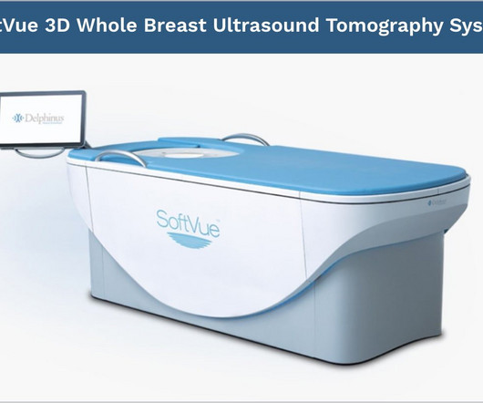
mtaschetta-millane Tue, 07/02/2024 - 09:50 July 2, 2024 — Delphinus Medical Technologies , a pioneering medicalimaging company that developed the SoftVue Breast Ultrasound Tomography (UST), announced today the publication of a study comparing mammography in conjunction with SoftVue UST vs mammography alone in women with dense breasts.
What’s the difference between Screening and Diagnostic Mammogram? During a diagnostic mammogram, the images are analyzed in real-time. During a diagnostic mammogram, the images are analyzed in real-time. Sometimes additional mammogramimages are taken.
Why Breast Density Matters in Cancer Screening Dense breast tissue affects screening in two key ways: Reduced Visibility : Dense tissue appears white on mammograms, as do tumors, making it harder to detect abnormalities. Inter-radiologist Variation : Assessments can vary up to 33% 1 when different radiologists interpret the same mammograms.
As we are turning the corner in the Fredericksburg region from COVID-19, MedicalImaging of Fredericksburg is pleased to resume scheduled appointments at all six of our locations. On May 4th, MedicalImaging of Fredericksburg will reopen all of its facilities for scheduled appointments.
MedicalImaging of Fredericksburg Ranked First Place in “Best of the Burg” 2019 Contest MedicalImaging of Fredericksburg (MIF) took first place in the 2019 “Best of the Burg” contest, earning the award for “Best MedicalImaging”.
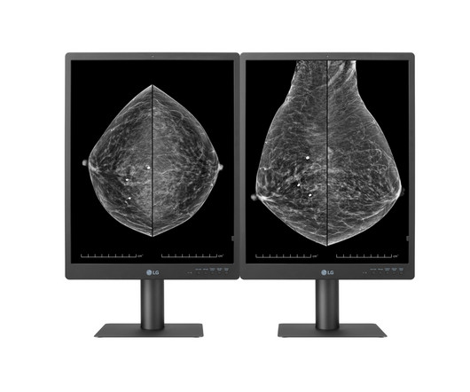
Suitable for reviewing various types of medicalimages, including breast MRIs, CT scans and ultrasound, the diagnostic monitor features an internal front sensor that removes the need for an external calibration device. Hu, LG Business Solutions USA’s head of medical displays.
However, this can make it a bit trickier to detect breast cancer with a standard mammogram. Your breast density is something your radiologist determines when they read your mammogram. This information is included in your mammogram results, so be sure to ask your doctor about your breast density.
Try as we might, good health practices tend to be neglected, especially regarding preventive health screenings like mammograms. Research shows that one in four women who should be getting regular mammograms don’t. Women with an average risk for breast cancer should begin mammogram screening every year starting at 40.
Her story is featured in an upcoming themed issue of the Journal of MedicalImaging and Radiation Sciences on the topic of specialized populations, published by Elsevier. Despite having a lump in her breast for several decades, she was unable to undergo mammograms due to the lack of accessible equipment and procedures.
milla1cf Tue, 06/18/2024 - 20:09 June 18, 2024 — Delphinus Medical Technologies , a pioneering medicalimaging company that developed the SoftVue Breast Tomographic Ultrasound, today announced that Elise Berman , M.D., has joined the company as medical director. area’s premier breast imaging practices. “We
Remember that breast self-exams are part of a comprehensive breast health routine, and regular clinical exams and mammograms are also important for early detection of breast cancer. In women who have dense breast tissue, additional imaging tests such as whole breast ultrasound or MRI may help to detect additional cancers.
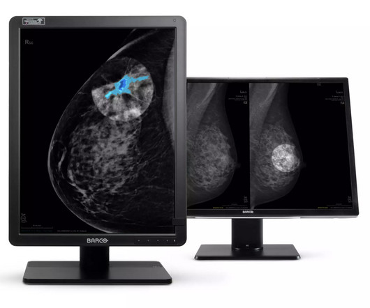
Combined with our displays’ excellent image quality, this tool offers a new layer of confidence to breast radiologists. DL Precise will be integrated with Barco display systems, enabling deployment across hundreds of medicalimaging viewers used by thousands of clinicians.
Depending on the diagnosis and severity of the patient’s condition, a physician may use a wide array of imaging tests to help determine the best cancer treatment. This method of imaging is most frequently used to look for cancer in the abdomen as well as guiding a biopsy in breast tissue.
The theoretical basis for ultrasound physics has been around since 1794, but it wasn’t until 1942, when Dr Karl Theodore Dussik in Austria transmitted an ultrasound beam through a human skull to view the brain, that ultrasound was first used in medicine. (13) This was a defining publication in the field of medicalultrasound. (14)
We organize all of the trending information in your field so you don't have to. Join 5,000 users and stay up to date on the latest articles your peers are reading.
You know about us, now we want to get to know you!
Let's personalize your content
Let's get even more personalized
We recognize your account from another site in our network, please click 'Send Email' below to continue with verifying your account and setting a password.


Let's personalize your content