This site uses cookies to improve your experience. To help us insure we adhere to various privacy regulations, please select your country/region of residence. If you do not select a country, we will assume you are from the United States. Select your Cookie Settings or view our Privacy Policy and Terms of Use.
Cookie Settings
Cookies and similar technologies are used on this website for proper function of the website, for tracking performance analytics and for marketing purposes. We and some of our third-party providers may use cookie data for various purposes. Please review the cookie settings below and choose your preference.
Used for the proper function of the website
Used for monitoring website traffic and interactions
Cookie Settings
Cookies and similar technologies are used on this website for proper function of the website, for tracking performance analytics and for marketing purposes. We and some of our third-party providers may use cookie data for various purposes. Please review the cookie settings below and choose your preference.
Strictly Necessary: Used for the proper function of the website
Performance/Analytics: Used for monitoring website traffic and interactions
Mammograms are a crucial diagnostic tool that helps doctors detect early signs of breast cancer and other breast-related issues. However, many patients have concerns about radiation exposure and the potential risks involved. Radiation exposure is controlled and minimized to ensure patient safety.
Previous research suggests that when the procedure is combined with hormonal therapy and radiation, patients can have nearly 100% of their tumors destroyed, Bryce noted. hormone therapy and radiation) therapies can have on this patient population. hormone therapy and radiation) therapies can have on this patient population.
A team led by Vivianne Freitas, MD, from the University of Toronto found that PEM performed comparably to MRI in breast cancer detection and could serve as a supplemental imaging method for evaluating dense breasts. This is where supplemental imaging such as MRI and molecular breast imaging come into play.
Amidst the battle against this disease, screening mammograms emerge as a crucial tool in early detection and effective treatment. In this blog, we delve into the significance of screening mammograms, their procedure, their benefits, and why they are essential for women’s health. What is a Screening Mammogram?
In an October 15 communication , the agency said it had received reports of electronic medical devices being damaged during CT scans due to radiation. Interference is when the radiation and the device electronics are incompatible, and the resulting damage causes the device to fail to work normally," the FDA said.
While this is an improvement over the prior recommendation to begin screening at age 50, it falls short of the current recommendation by the Society of Breast Imaging and the American College of Radiology to obtain annual mammograms beginning at age 40. Concern for radiation risk due to annual screening is really of no concern!
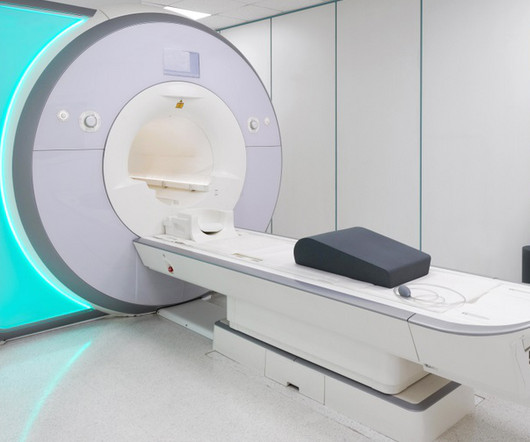
That changed in 2023, when Angie underwent a mammogram and breast ultrasound at a Midstate Radiology Associates location offering CARE. Based on Angie’s risk score, she qualified for a breast MRI, a diagnostic tool capable of detecting cancers that mammograms and ultrasounds might miss.
If you’re in your 30’s, you may be wondering if it’s time to add a yearly mammogram to your healthcare routine. When Does the American Cancer Society Recommend Starting Mammograms? Continue to get mammograms if you’re in good health and expect to live an additional 10 years. Should I Get a Mammogram in My 20s or 30s?
Since almost half of the screening population has dense breasts, many of these patients require additional breast imaging, often with MRI , after mammography. Low-dose positron emission mammography (PEM) is a novel molecular imaging technique that provides improved diagnostic performance at a radiation dose comparable to that of mammography.
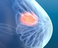
The earlier breast cancer is detected through diagnostic imaging, the better chance there is for successful treatment with surgery, radiation therapy, or chemotherapy. Your physician will do a physical examination, sometimes including mammography, as well as ask questions about your medical history and lifestyle. this year alone.
For dense-breasted patients requiring supplemental imaging, MRI remains a valuable option that is not limited by breast density and is shown to be more sensitive than mammography at finding breast cancer. J Med Imaging Radiat Sci. The shortage of radiographers: A global crisis in healthcare. 2023 Oct 19:S1939-8654(23)01877-5.
However, this can make it a bit trickier to detect breast cancer with a standard mammogram. Your breast density is something your radiologist determines when they read your mammogram. This information is included in your mammogram results, so be sure to ask your doctor about your breast density.
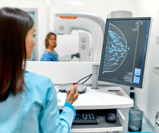
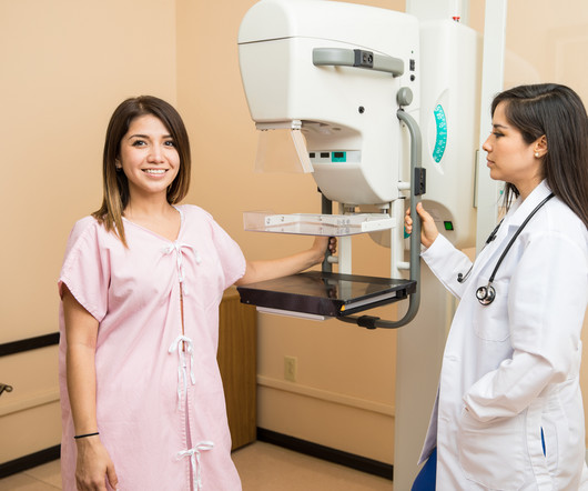
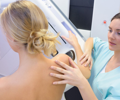
Routine mammograms and other recommended breast screenings can help improve patient outcomes by paving the way for early detection. Types of Breast Screening MammogramMammograms by and far account for the largest number of routine breast screenings. When getting a mammogram, the patient stands in front of the machine.
Try as we might, good health practices tend to be neglected, especially regarding preventive health screenings like mammograms. Research shows that one in four women who should be getting regular mammograms don’t. Women with an average risk for breast cancer should begin mammogram screening every year starting at 40.
MRI-Scan-Teleradiology Introduction: Mammography is not just a diagnostic tool; it’s a mastery of precision and compassion in breast imaging. This blog is a journey through the world of mammography, shedding light on the importance of precision and compassion in breast health.
The compression results in the reduced thickness of the breast and the radiation to the breast are decreased. Hence, mammograms carried out anywhere can now be viewed by expert breast radiologists in any part of the world thanks to teleradiology. If the breast is dense on the mammogram, an ultrasound must also be carried out.
What’s more, the USPSTF concluded that there was insufficient evidence to recommend supplemental screening with MRI or ultrasound in women, regardless of breast density. Furthermore, high-risk women who desire supplemental screening -- but cannot undergo MRI -- should consider contrast-enhanced mammography, according to the ACR.
Such harms may include unnecessary biopsy, additional radiation exposure with more imaging, and patient anxiety. Anxiety from an inconclusive mammogram result or false positive is brief, with no lasting health effects. He also wrote that overdiagnosis can be harmful to women.
3) The British Röntgen Society (the first radiology society) was founded in 1897, and many further studies on X-ray usage and the effects of radiation were performed over the following years. (3) Their work gave rise to the modern MRI scanners we use today. 3) This is what is known as tomography. 1971 Mar 19;171(3976):1151-3.
We organize all of the trending information in your field so you don't have to. Join 5,000 users and stay up to date on the latest articles your peers are reading.
You know about us, now we want to get to know you!
Let's personalize your content
Let's get even more personalized
We recognize your account from another site in our network, please click 'Send Email' below to continue with verifying your account and setting a password.


Let's personalize your content