This site uses cookies to improve your experience. To help us insure we adhere to various privacy regulations, please select your country/region of residence. If you do not select a country, we will assume you are from the United States. Select your Cookie Settings or view our Privacy Policy and Terms of Use.
Cookie Settings
Cookies and similar technologies are used on this website for proper function of the website, for tracking performance analytics and for marketing purposes. We and some of our third-party providers may use cookie data for various purposes. Please review the cookie settings below and choose your preference.
Used for the proper function of the website
Used for monitoring website traffic and interactions
Cookie Settings
Cookies and similar technologies are used on this website for proper function of the website, for tracking performance analytics and for marketing purposes. We and some of our third-party providers may use cookie data for various purposes. Please review the cookie settings below and choose your preference.
Strictly Necessary: Used for the proper function of the website
Performance/Analytics: Used for monitoring website traffic and interactions
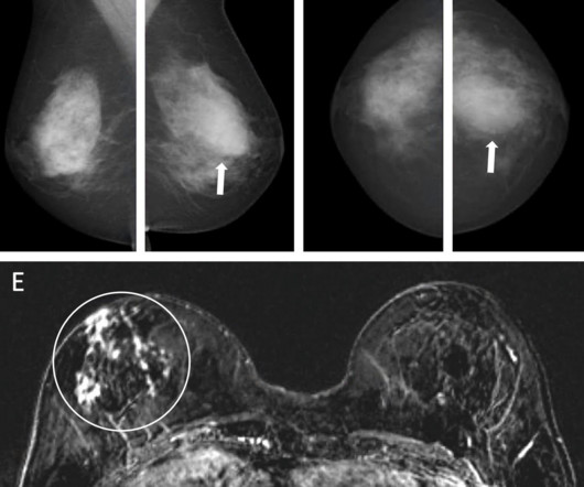
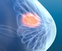
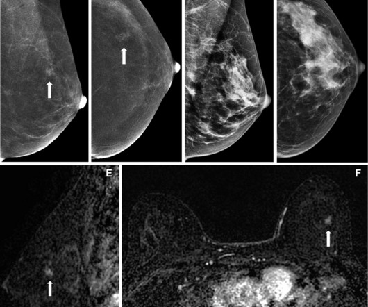
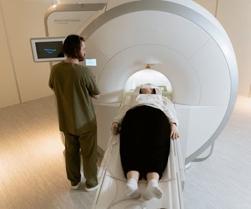
Using AI with mammography can help select women at high risk of breast cancer for supplemental MRI, according to research published February 4 in Radiology. However, access to MRI can be limited for some women. E) Concurrent axial subtraction MRI scan shows a large, diffuse, invasive lobular cancer (circle) in the right breast. (E)
million mammograms were performed in the U.S. Although mammogram is the most widely used screening modality, a known problem is that 9.5% Starting with mammography images, the work is important for finding ways to alleviate the region's radiologist workforce shortage and staffing pressures, according to Imperial College London.
An act making mammograms more accessible in Illinois is now in lawmakers’ hands. Coverage for molecular breast imaging will be required and, in those cases where it is not already covered, breast MRI will also be covered.
Abbreviated MRI may be suitable for screening women with dense breasts, a study published May 22 in the American Journal of Roentgenology found. Radiologists and ordering clinicians, alike, may use these results as evidence of the value of abbreviated MRI beyond the first round of screening in this population," Edmonds told AuntMinnie.com.
A radiologist’s perception when viewing a complex MR image may be akin to a Major League Baseball (MLB) batter reading the stitches on a fastball, according to researchers exploring exactly how diagnostic interpretations are made. They found that some observers performed better than expected for their rank and years of experience.
A team led by Su Min Ha, MD, PhD, from Seoul National University Hospital in South Korea reported that AI by itself achieved a higher performance than radiologists with no AI help when it came to detecting contralateral breast cancer in women treated with unilateral mastectomy. The radiologists had 10 and 17 years of experience, respectively.
A team led by Vivianne Freitas, MD, from the University of Toronto found that PEM performed comparably to MRI in breast cancer detection and could serve as a supplemental imaging method for evaluating dense breasts. This is where supplemental imaging such as MRI and molecular breast imaging come into play.
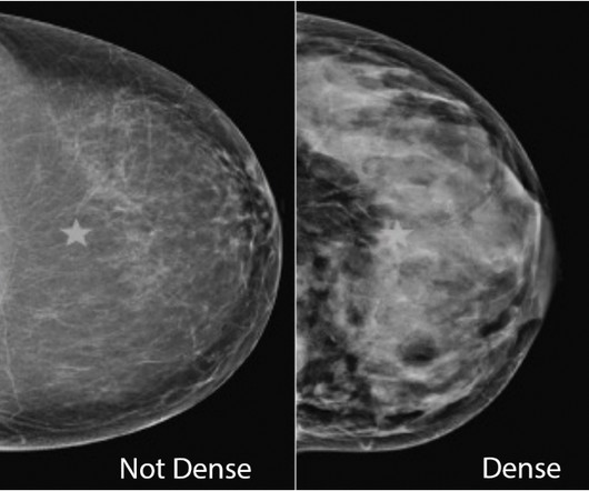
Breast density assessment is important not only for the patients health, but it also has reimbursement implications for radiologists. Breast density can often obscure lesions on conventional x-ray mammography, and so other screening modalities such as MRI or ultrasound are often recommended for follow-up.
As more people get vaccinated for COVID-19, radiologists must be familiar with how the vaccine may affect imaging results, wrote Shabnam Mortazavi, MD, of the University of California, Los Angeles. insert table here) (Above) 55-year-old woman who underwent screening mammogram and ultrasound seven days after first COVID-19 vaccination dose.
The finding suggests the technique may provide a new treatment path for women who are not candidates for lumpectomy, or surgical removal, noted Yolanda Bryce, MD, an interventional radiologist at Memorial Sloan Kettering Cancer Center in New York City, and senior author of the study.
Across the United States, radiologist shortages are creating a ripple effect that many patients never seeuntil theyre left waiting. Another waited three months for mammogram findings. Neiman Health Policy Institute, the radiologist shortage is expected to continue through 2055 if action isn’t taken.
A recent Australian study criticizing narratives surrounding breast density notification has drawn mixed reactions from radiologists and advocates of breast cancer screening. Radiologists have studied and continue to research how breast density plays into breast cancer risk. The law goes into effect September 10, 2024.
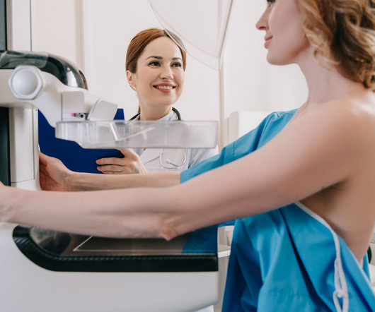
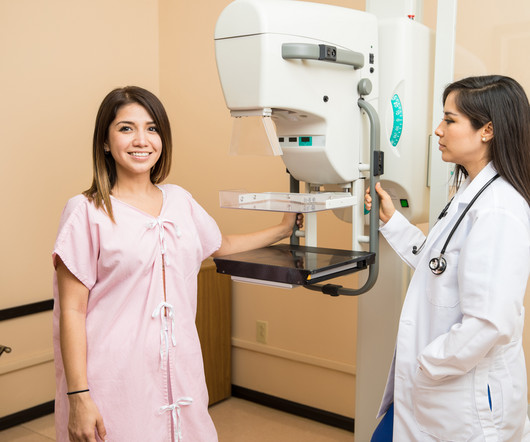
Amidst the battle against this disease, screening mammograms emerge as a crucial tool in early detection and effective treatment. In this blog, we delve into the significance of screening mammograms, their procedure, their benefits, and why they are essential for women’s health. What is a Screening Mammogram?
In testing, the score outperformed traditional mammographic density measurements in flagging patients for supplemental breast imaging following a negative screening mammogram. Moreover, this risk categorization might also prompt an additional image review by radiologists, particularly when informed of a high AISmartDensity score.”
Why Breast Density Matters in Cancer Screening Dense breast tissue affects screening in two key ways: Reduced Visibility : Dense tissue appears white on mammograms, as do tumors, making it harder to detect abnormalities. Inter-radiologist Variation : Assessments can vary up to 33% 1 when different radiologists interpret the same mammograms.
New AI integrations are further elevating the impact of this technology, not only by acting as a valuable second set of eyes for radiologists but also by providing standardized breast density assessments. As the technology matures and algorithms are further refined, radiologists are gaining greater confidence in the specificity of results.
A team led by Joao Horvat, MD, from the Memorial Sloan Kettering Cancer Center in New York found that CEM depicted 90% of breast cancers compared with 10% on low-energy mammograms alone and 50% on low-energymammogramswith whole-breast ultrasound. Horvat and co-authors investigated whether the same trend goes for CEM.
Finally, two expert radiologists isolated the regions of interest of the primary tumor from other areas of breast tissue. From each patient, the model extracted 4,096 features from mammograms and selected six related features according to the relationship between tumor size determination and lymph node metastasis. International.
If you’re in your 30’s, you may be wondering if it’s time to add a yearly mammogram to your healthcare routine. When Does the American Cancer Society Recommend Starting Mammograms? Continue to get mammograms if you’re in good health and expect to live an additional 10 years. Should I Get a Mammogram in My 20s or 30s?
Techniques such as mammography, low-dose computed tomography (LDCT), and magnetic resonance imaging (MRI) are instrumental in identifying cancers like breast, lung, and prostate in their nascent stages. This technology mitigates the challenges posed by a shortage of on-site radiologists and enhances the quality of care in remote areas.
Further, the task force incorrectly concludes there is “inadequate” evidence to support adding screening MRI (or ultrasound if MRI is not possible) after a mammogram for many women with dense breasts. In women with the densest breasts, about 40% of cancers are missed on a mammogram.
Since almost half of the screening population has dense breasts, many of these patients require additional breast imaging, often with MRI , after mammography. Two breast radiologists reviewed PEM images taken one and four hours post 18 F-FDG injection and correlated the findings with lab results.
“In recent years, AI has been studied for the purpose of diagnosing breast cancer earlier by automatically detecting breast cancers in mammograms and measuring the risk of future breast cancer.” Diagnostic AI models are trained to detect suspicious lesions on mammograms and are well suited to estimate short-term breast cancer risk.
However, this can make it a bit trickier to detect breast cancer with a standard mammogram. Your breast density is something your radiologist determines when they read your mammogram. This information is included in your mammogram results, so be sure to ask your doctor about your breast density.
1) After a mammogram, other screening tests such as ultrasound, and especially MRI, find additional cancers in dense breasts. Women should discuss density and other risk factors with their radiologist or health care provider. Detecting cancers missed by mammography can improve outcomes. Br J Radiol. 2012 Nov;85(1019):1465-70.
Women may feel as though they are constantly hearing about the importance of regular mammograms. Breast Density and Optimal Screening Outcomes When advised to get a mammogram, most women do not think about the type of breasts they have. Unless you have implants, then you may need an MRI. Breasts are breasts, right?
The approval expands upon Bayer's focus on breast imaging, with a portfolio that also includes Gadavist (gadobutrol) injection, a gadolinium-based contrast agent approved for use with MRI ( Magnetic Resonance Imaging ) to assess the presence and extent of malignant breast disease in adult patients. Breast Density on a Mammogram.
I see it acting like a bridge between the referring healthcare professional and the expert radiologist — stepping in as a trained consultant to recommend the right imaging test at the point of care, without delay. It might be an MRI, an ultrasound, a mammogram, or another imaging test. answered an average of 88.9%
Try as we might, good health practices tend to be neglected, especially regarding preventive health screenings like mammograms. Research shows that one in four women who should be getting regular mammograms don’t. Women with an average risk for breast cancer should begin mammogram screening every year starting at 40.
Women with dense breasts are BOTH more likely to develop breast cancer and more likely to have that cancer missed on a mammogram [5] Fig. 1 – Cancer on a mammogram of a fatty vs a dense breast What is Dense Breast Tissue? Breast density is determined through a mammogram and described as one of four categories (Fig.
Medical Imaging of Fredericksburg (MIF) is a partnership between Radiologic Associates of Fredericksburg and Mary Washington Healthcare, whose Board Certified, Fellowship Trained Radiologists serve the community in five counties in Virginia with convenient access to the highest level of care for the greatest value in the region.
In an effort to help our clients address the backlog created by this dramatic decrease earlier this year, Direct Radiology now offers dedicated women’s imaging radiologists as part of its full-service teleradiology practice. Why is the timing critical? Experts indicate that this year is no exception to that trend. “As
MRI-Scan-Teleradiology Introduction: Mammography is not just a diagnostic tool; it’s a mastery of precision and compassion in breast imaging. This blog is a journey through the world of mammography, shedding light on the importance of precision and compassion in breast health.
A couple of weeks later, I received a phone call at 9:00pm from a radiologist who was starting her own breast center. This radiologist had just returned from Sweden, where she completed a breast fellowship with Dr. László Tabár, a huge believer in comprehensive breast care. She was passionate and excited about her future plans.
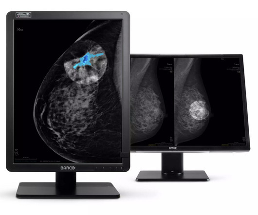
Combined with our displays’ excellent image quality, this tool offers a new layer of confidence to breast radiologists. DL Precise will be integrated with Barco display systems, enabling deployment across hundreds of medical imaging viewers used by thousands of clinicians.
Whether you’re looking for a second set of eyes for complex cases or want to ensure the highest level of diagnostic accuracy, our team of board-certified radiologists—with subspecialties in areas such as neuroradiology, musculoskeletal imaging, and oncology—are ready to assist. Why Choose a Teleradiology Partner for Second Opinions?
A) Mammogram MLO view. Mammogram CC view. A mammogram demonstrated focal asymmetries involving most of the anterior and mid right breast with diffuse skin thickening, trabecular coarsening, increased overall density, and enlarged right axillary lymph nodes. What is the diagnosis? Xray of the Week Figure 1. Surg Clin North Am.
Hence, mammograms carried out anywhere can now be viewed by expert breast radiologists in any part of the world thanks to teleradiology. If the breast is dense on the mammogram, an ultrasound must also be carried out. Depending on the risk, at times MRI breast is started annually starting at 25 years and mammography at 35.
There are several types of imaging tests that physicians use to detect cancer in patients: X-Ray, Computed Tomography (CT), Magnetic Resonance Imaging (MRI), Ultrasound (US), Nuclear Medicine, and Positron Emission Tomography (PET). Our radiologists also consult closely with referring physicians on many cancer-related cases.
All of the annual scheduled services such as mammograms can now be scheduled, as well as imaging prescribed by physicians for the care of their patients. Patients may also schedule mammograms directly at our facilities in these same locations. Christopher Newman, Chief Medical Officer of Mary Washington Healthcare.
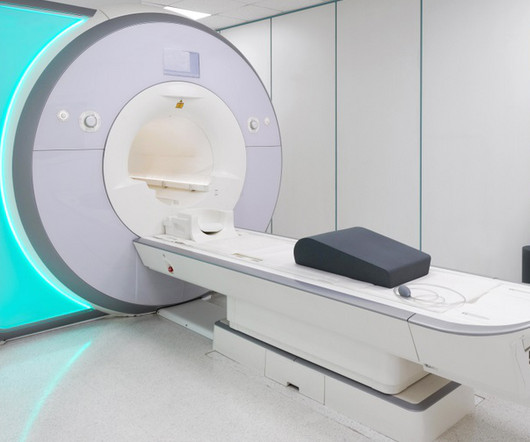
To mitigate the time challenges typically faced when reporting tomosynthesis images, advanced imaging technology can reconstruct high-resolution tomosynthesis slices which results in a reduction in radiologist reading time. What Is a Breast MRI? Radiology: Volume 286: Number 3—March 2018 [4] Cancer.org. Breast Cancer Screening.
Radiologists often have had little experience or training in item-writing, and, as a group, they are incredibly busy. An MCQ that asks the learner to recognize benign dermal calcifications on a mammogram does not test the learner’s problem-solving ability or ability to communicate the findings to a patient.
What’s more, the USPSTF concluded that there was insufficient evidence to recommend supplemental screening with MRI or ultrasound in women, regardless of breast density. Furthermore, high-risk women who desire supplemental screening -- but cannot undergo MRI -- should consider contrast-enhanced mammography, according to the ACR.
We organize all of the trending information in your field so you don't have to. Join 5,000 users and stay up to date on the latest articles your peers are reading.
You know about us, now we want to get to know you!
Let's personalize your content
Let's get even more personalized
We recognize your account from another site in our network, please click 'Send Email' below to continue with verifying your account and setting a password.


Let's personalize your content