This site uses cookies to improve your experience. To help us insure we adhere to various privacy regulations, please select your country/region of residence. If you do not select a country, we will assume you are from the United States. Select your Cookie Settings or view our Privacy Policy and Terms of Use.
Cookie Settings
Cookies and similar technologies are used on this website for proper function of the website, for tracking performance analytics and for marketing purposes. We and some of our third-party providers may use cookie data for various purposes. Please review the cookie settings below and choose your preference.
Used for the proper function of the website
Used for monitoring website traffic and interactions
Cookie Settings
Cookies and similar technologies are used on this website for proper function of the website, for tracking performance analytics and for marketing purposes. We and some of our third-party providers may use cookie data for various purposes. Please review the cookie settings below and choose your preference.
Strictly Necessary: Used for the proper function of the website
Performance/Analytics: Used for monitoring website traffic and interactions
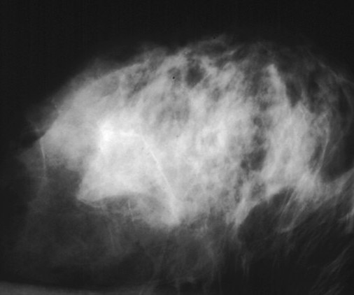
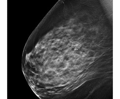
Why Breast Density Matters in Cancer Screening Dense breast tissue affects screening in two key ways: Reduced Visibility : Dense tissue appears white on mammograms, as do tumors, making it harder to detect abnormalities. mammography facilities must provide patients with information about their breast density.
Because thorough validation of AI models is crucial for effective patientcare, and as the U.S. AI has shown promise for streamlining radiologists' workflows, from distinguishing normal from abnormal mammograms or chest x-rays to helping predict cardiac disease risk.
New AI integrations are further elevating the impact of this technology, not only by acting as a valuable second set of eyes for radiologists but also by providing standardized breast density assessments. vi Investigations continue of this newer imaging modality, which has the potential to positively benefit patients with dense breasts.
This technology mitigates the challenges posed by a shortage of on-site radiologists and enhances the quality of care in remote areas. A report from Healthcare IT News highlights how teleradiology enables radiologists to interpret scans remotely, increasing flexibility in work schedules and expanding access to specialized expertise.
With AI-mediated cyberattacks on the rise, radiologists can find it challenging to balance prompt patient access to diagnostic imaging while safeguarding sensitive healthcare data. Dhaval Shah. With growing radiology practices and outpatient imaging facilities, we will see an increase in outsourced diagnostic imaging.
Maybe you have recently moved, changed doctors, or have new insurance and now, are going to a different imaging center for your annual mammogram. Either way, the new imaging center will very often request that you provide the images from your previous mammograms. In this case, the radiologist may recommend a diagnostic mammogram.
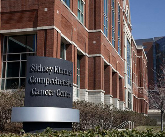
christine.book Wed, 03/13/2024 - 12:18 March 13, 2024 — ScreenPoint Medical has announced that its industry leading Transpara Breast AI is available to improve cancer detection for Johns Hopkins Medicine breast cancer screening patients. Transpara is designed to work concurrently with radiologists.
By reducing overhead costs associated with maintaining in-house radiologists, rural hospitals can ensure uninterrupted imaging services without compromising care quality. Radiologists must inform patients if they have dense breast tissue, a factor that can obscure mammogram results and increase cancer risks.
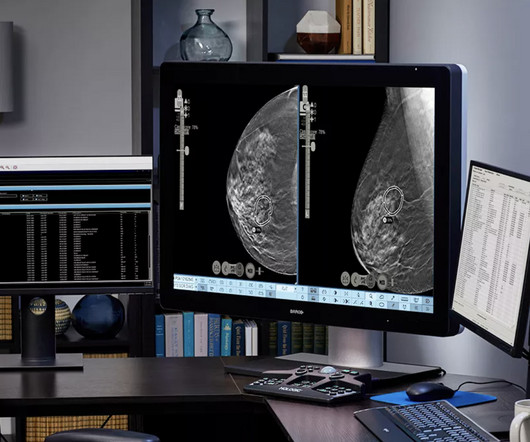
With the latest high-end medical display technology from LG, physicians can analyze radiological and mammogram images with complete confidence. With the LG Digital X-Ray Detector, radiologists and physicians can examine clear x-ray images, fast. LG Digital X-Ray Detector (Model 14HQ701G) The LG Digital X-Ray Detector.
This technology helps to augment radiologists by providing a concurrent read of screening and diagnostic mammograms, often enhancing early detection of breast cancer. “We We are thrilled to collaborate with US Radiology to bring our advanced AI-powered mammography solutions to a broader patient base,” said Dana Brown, CEO of iCAD, Inc.
With an X-ray, Ultrasound, Mammogram and CT scan at our disposal, we needed to have a radiologist who could read all these modalities, and give us results in the shortest time possible to enable us to give the best medical care possible” she says. It is like having a radiologist on site with each subspecialty represented!”
In recent years, artificial intelligence (AI) has made remarkable strides in revolutionizing the landscape of the medical field, offering unprecedented opportunities for enhanced patientcare, diagnosis, and treatment. In the United States alone, it is estimated that around 40 million mammograms were performed each year.
“The conventional double-reading workflow utilized by most countries, where mammograms are assessed by two separate radiologists, has become increasingly challenging due to the scarcity of radiologists worldwide. But one company can’t make this a reality alone,” said Greg Corrado , Ph.D., Head of Health AI, Google.
“iCAD's Breast AI Suite is the only solution on the market today offering a complete portfolio of breast cancer detection, density assessment, and risk evaluation solutions that are clinically proven to increase cancer detection, assist radiologists in assessing short-term cancer risk, improve workflow, and enhance patientcare.
I see it acting like a bridge between the referring healthcare professional and the expert radiologist — stepping in as a trained consultant to recommend the right imaging test at the point of care, without delay. It might be an MRI, an ultrasound, a mammogram, or another imaging test. answered an average of 88.9%
In a significant move to enhance breast cancer detection and patientcare, the U.S. Effective as of September 2024, this new amendment mandates that all mammography facilities include information about breast density in their mammography reports and results letters to patients. What Does This Mean for Patients?
The pioneer behind 3D mammography , Hologic will present new data that showcase how next-generation deep-learning solutions can assist with breast cancer detection and improve workflow for radiologists. CET, is designed to provide radiologists with an understanding of how CEM can be effectively implemented into their daily practice.
most patients don’t like having mammograms! Here are some ideas, especially for October, but some of which can be practiced throughout the year: Kindness, Compassion and Empathy Being a woman gives us the unique opportunity to relate to our patients. Communication Communication should be correct, concise and caring.
When it comes to accurate diagnoses and effective patientcare, getting a second opinion on imaging results can make all the difference. Mammography and Breast Imaging Given the sensitivity and potential impact of findings on patientcare, a second opinion can confirm initial readings and prevent over- or under-treatment.
As I was looking over the menu, I thought to myself, “Wouldn’t it be nice if we could ‘order up’ our patients!!” OK, I know, I know.but it is never far from my mind, or yours, as I suspect that all mammographers look at people we see/meet as potential mammogrampatients! And aren’t we glad that radiologists are so thorough??!!
Clinical studies have demonstrated that the use of iCAD’s ProFound AI solutions can significantly improve reading sensitivity and specificity and reduce reading times, thus enhancing clinical decision support and workflow efficiency for radiologists. milla1cf Tue, 05/28/2024 - 07:00 May 28, 2024 — iCAD, Inc.,
Board Certified radiologists, we are committed to bridging the gap by offering accurate and timely readings. Whether it’s X-rays, MRIs, mammograms , CT scans, or other subspecialty , our streamlined process ensures swift delivery of results without compromising on quality.
Burnout is rampant and I cannot help but believe that as a result, patientcare may be compromised. and then, in turn, patientcare, I am never one to sit back and say, "Well, that’s a bummer." I have always maintained the theory that those who do not like to perform mammograms should not do so.
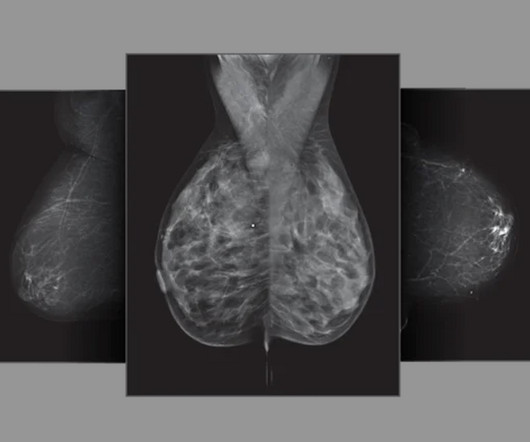
More accurate and efficient detection is instrumental for better patient outcomes. 3D Mammography systems have become smarter, bringing breast cancer diagnosis to a new level, improving cancer detection accuracy, optimising workflow, and supporting personalised patientcare [1].
There are several barriers that still need to be addressed from a radiologist, facility, and patient standpoint. How someone can extrapolate findings from a study conducted overseas with less than 100 participants in a publicly funded healthcare system and present it as a universal truth to over 49,000 radiologists in the U.S.
28, 2024 — Rezolut, LLC recently debuted its latest offering for patients during their annual mammogram, SecondReadAI powered by Lunit. Leveraging deep learning techniques, the system analyzes mammography images, providing radiologists with valuable insights and supporting them in their diagnostic process.
The study’s limitations include the potential misclassification of diagnostic mammograms as screening and the inability to adjust for certain breast cancer risk factors. The post Breast Cancer Awareness Month Kicks Off Now: The Latest in Breast Cancer Studies first appeared on Vesta Teleradiology.
On behalf of ACR — representing more than 41,000 diagnostic radiologists, interventional radiologists, radiation oncologists, nuclear medicine physicians and medical physicists, he urged the Subcommittee to provide at least $50.9 billion to the NIH for fiscal year (FY) 2024, an increase of $3.5
A clear blueprint for standardized protocols is not just another checkbox for compliance, but can serve as a solid foundation for high quality patientcare and seamless workflow efficiency for all team members. The likelihood of a radiologist being the defendant in at least one lawsuit is 50% by age 60. A study by Shah et al.
IR came to life through the combination of the creative thinking and technical skills of diagnostic radiologists and angiogiographers. (11) 11) Charles Dotter, a vascular radiologist and commonly known as ‘The Father of Interventional Radiology’, invented angioplasty. (12) 15) Radiologists needed a common means for sharing images.
We organize all of the trending information in your field so you don't have to. Join 5,000 users and stay up to date on the latest articles your peers are reading.
You know about us, now we want to get to know you!
Let's personalize your content
Let's get even more personalized
We recognize your account from another site in our network, please click 'Send Email' below to continue with verifying your account and setting a password.


Let's personalize your content