This site uses cookies to improve your experience. To help us insure we adhere to various privacy regulations, please select your country/region of residence. If you do not select a country, we will assume you are from the United States. Select your Cookie Settings or view our Privacy Policy and Terms of Use.
Cookie Settings
Cookies and similar technologies are used on this website for proper function of the website, for tracking performance analytics and for marketing purposes. We and some of our third-party providers may use cookie data for various purposes. Please review the cookie settings below and choose your preference.
Used for the proper function of the website
Used for monitoring website traffic and interactions
Cookie Settings
Cookies and similar technologies are used on this website for proper function of the website, for tracking performance analytics and for marketing purposes. We and some of our third-party providers may use cookie data for various purposes. Please review the cookie settings below and choose your preference.
Strictly Necessary: Used for the proper function of the website
Performance/Analytics: Used for monitoring website traffic and interactions
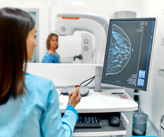
Mammograms are a crucial diagnostic tool that helps doctors detect early signs of breast cancer and other breast-related issues. However, many patients have concerns about radiation exposure and the potential risks involved. As the leading diagnostic imaging radiology center in El Paso, patientcare and safety are our top priorities.
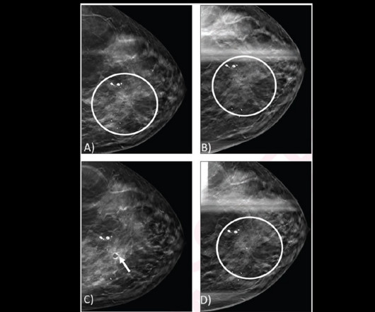
Researchers led by Derek Nguyen, MD, from Duke University in Durham, NC, found that for architectural distortions found by DBT alone with no ultrasound correlate, the malignancy rate was 0% for distortions without atypia versus 20% for distortions with atypia on core needle biopsy. No ultrasound correlate was identified (not shown).
Why Breast Density Matters in Cancer Screening Dense breast tissue affects screening in two key ways: Reduced Visibility : Dense tissue appears white on mammograms, as do tumors, making it harder to detect abnormalities. Inter-radiologist Variation : Assessments can vary up to 33% 1 when different radiologists interpret the same mammograms.
For dense-breasted patients requiring supplemental imaging, MRI remains a valuable option that is not limited by breast density and is shown to be more sensitive than mammography at finding breast cancer. vi Investigations continue of this newer imaging modality, which has the potential to positively benefit patients with dense breasts.
Maybe you have recently moved, changed doctors, or have new insurance and now, are going to a different imaging center for your annual mammogram. Either way, the new imaging center will very often request that you provide the images from your previous mammograms. In this case, the radiologist may recommend a diagnostic mammogram.
They include digital breast exams (DBE), clinical breast exams (CBE), self-examination, 3D mammograms, MRI and CT scans, ultrasound, blood tests, diagnostic imaging, and biopsies. Our entire staff is committed to exceptional patientcare. It is up to you and your doctor to decide what is right for you.
With an X-ray, Ultrasound, Mammogram and CT scan at our disposal, we needed to have a radiologist who could read all these modalities, and give us results in the shortest time possible to enable us to give the best medical care possible” she says. MHRG devised a 24/7 remote radiology solution for the Clinic.
“iCAD's Breast AI Suite is the only solution on the market today offering a complete portfolio of breast cancer detection, density assessment, and risk evaluation solutions that are clinically proven to increase cancer detection, assist radiologists in assessing short-term cancer risk, improve workflow, and enhance patientcare.
In a significant move to enhance breast cancer detection and patientcare, the U.S. Effective as of September 2024, this new amendment mandates that all mammography facilities include information about breast density in their mammography reports and results letters to patients. What Does This Mean for Patients?
When a primary care doctor orders specialized testing, say for a patient who complains of breast pain, they may not know the best imaging test to choose. It might be an MRI, an ultrasound, a mammogram, or another imaging test. For more information: massgeneralbrigham.org Wednesday, July 5, 2023 - 19:42
When it comes to accurate diagnoses and effective patientcare, getting a second opinion on imaging results can make all the difference. Mammography and Breast Imaging Given the sensitivity and potential impact of findings on patientcare, a second opinion can confirm initial readings and prevent over- or under-treatment.
As I was looking over the menu, I thought to myself, “Wouldn’t it be nice if we could ‘order up’ our patients!!” OK, I know, I know.but it is never far from my mind, or yours, as I suspect that all mammographers look at people we see/meet as potential mammogrampatients! Answer questions honestly, without scaring the patient!
The theoretical basis for ultrasound physics has been around since 1794, but it wasn’t until 1942, when Dr Karl Theodore Dussik in Austria transmitted an ultrasound beam through a human skull to view the brain, that ultrasound was first used in medicine. (13) This was a defining publication in the field of medical ultrasound. (14)
We organize all of the trending information in your field so you don't have to. Join 5,000 users and stay up to date on the latest articles your peers are reading.
You know about us, now we want to get to know you!
Let's personalize your content
Let's get even more personalized
We recognize your account from another site in our network, please click 'Send Email' below to continue with verifying your account and setting a password.


Let's personalize your content