This site uses cookies to improve your experience. To help us insure we adhere to various privacy regulations, please select your country/region of residence. If you do not select a country, we will assume you are from the United States. Select your Cookie Settings or view our Privacy Policy and Terms of Use.
Cookie Settings
Cookies and similar technologies are used on this website for proper function of the website, for tracking performance analytics and for marketing purposes. We and some of our third-party providers may use cookie data for various purposes. Please review the cookie settings below and choose your preference.
Used for the proper function of the website
Used for monitoring website traffic and interactions
Cookie Settings
Cookies and similar technologies are used on this website for proper function of the website, for tracking performance analytics and for marketing purposes. We and some of our third-party providers may use cookie data for various purposes. Please review the cookie settings below and choose your preference.
Strictly Necessary: Used for the proper function of the website
Performance/Analytics: Used for monitoring website traffic and interactions
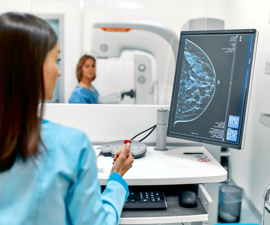
Mammograms are a crucial diagnostic tool that helps doctors detect early signs of breast cancer and other breast-related issues. The truth is mammograms are generally safe when used properly, and the amount of radiation you’re exposed to is minimal. Contact us online or call (915) 225-2480 to learn more.
Combining full-field digital mammography with ultrasound tomography can improve breast cancer detection and be helpful in imaging dense breasts, according to research published June 18 in Radiology. Our study suggests ultrasound tomography can be another supplemental screening tool,” Yamashita and colleagues wrote.
million mammograms were performed in the U.S. Although mammogram is the most widely used screening modality, a known problem is that 9.5% As is common in Europe, NHS currently uses a two-radiologist reader assessment of breast mammograms, three when there is disagreement. Just over 40.5
Gaps in coverage result in individuals paying anywhere from $234 for a follow-up diagnostic mammogram to over $1,000 for a breast MRI, Komen estimates.
It consists of a dual-energy digital mammogram performed with iodinated contrast and offers the same anatomic information as a digital mammogram. Reader 2 90% 93% p = 0.06 "Recombined images had better specificity compared to low energy in combination with ultrasound," the authors concluded. Reader 2 42% 53% p = 0.04
Maybe you recently decided to try the best 3D mammogram experience in El Paso, have recently moved, changed doctors, or acquired new insurance, and are now going to our imaging center for your annual mammogram. In this case, the radiologist may recommend a diagnostic mammogram. So, why are these prior images so important?
ChatGPT demonstrates modest accuracy when assigning BI-RADS scores for mammograms and breast ultrasound exams, according to research published October 30 in Clinical Imaging. Finally, both models achieved higher accuracy for mammograms at 67.6% compared with 55.6 % for ultrasound images. and 0.68, respectively.
Mortazavi and colleagues evaluated data from 23 women who presented with axillary adenopathy on mammography, breast ultrasound, or breast MRI after being vaccinated for COVID-19 between December 2020 and February 2021. Ultrasound from diagnostic work-up performed seven days later showed no change in lymph node size.
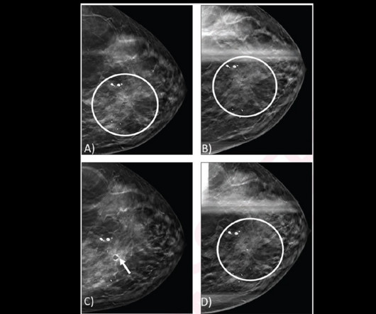
However, the researchers noted a lack of data on interpreting surveillance mammograms in women with a personal history of breast cancer. A) Left craniocaudal and (B) mediolateral oblique mammograms assessed as benign. (C) AI continues to show promise in improving screening mammography interpretation. years after right mastectomy. (A)
Breast density can often obscure lesions on conventional x-ray mammography, and so other screening modalities such as MRI or ultrasound are often recommended for follow-up. CEM is faster and less costly than MRI and can often be used as a follow-up to an abnormal screening mammogram when it is clinically appropriate.
Cryoablation uses imaging guidance typically with ultrasound or CT to locate tumors. Follow-up imaging was performed after the procedure by mammogram, ultrasound, or in some cases contrast-enhanced mammogram or MRI, based on patient eligibility and preference.
Amidst the battle against this disease, screening mammograms emerge as a crucial tool in early detection and effective treatment. In this blog, we delve into the significance of screening mammograms, their procedure, their benefits, and why they are essential for women’s health. What is a Screening Mammogram?
Previous studies have demonstrated that dense breast tissue masks breast cancers on mammography, and that supplemental imaging such as ultrasound and MRI confirms suspicious findings within dense tissue. Radiologists have studied and continue to research how breast density plays into breast cancer risk. The study can be found here.
While services for breast and lung cancer screening were temporarily halted, imagers in x-ray, lung ultrasound, and PET/CT were busy examining patients who presented with COVID-19. Her team employed a tracking mechanism for patients who were due for their mammograms once screening operations resumed.
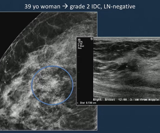
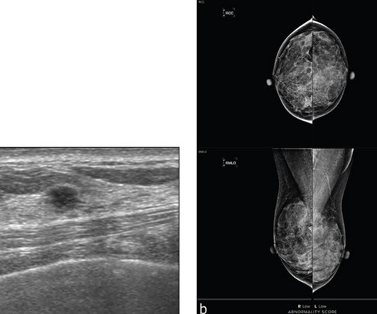
The retrospective study included data collected between 2007 and 2019 from 4,150 women in this age range who underwent 4,448 screening mammograms. Images of a 39-year-old woman shows a grade 2 invasive ductal carcinoma on mammography, confirmed by supplementary ultrasound.
Gina Curry (D-Delaware) and would eliminate costs for women for supplemental imaging such as breast MRIs and ultrasounds. When a mammogram reveals an abnormality or an individual is at a higher risk of breast cancer, diagnostic and supplemental imaging is required to determine if the patient needs a biopsy. Komen, in a news release.
The reduction for the bilateral mammogram 77066 was 1.36%, reflecting an increase in RVU valuation that somewhat offsets the conversion factor cut. Effect on professional component reimbursement The single-view chest x-ray 71045 professional fee was cut 5.55%.
The USPSTF also said that there was insufficient evidence to recommend supplemental screening with MRI or ultrasound in women, regardless of breast density. If you’re in good health, keep having mammograms as long as you’re allowed to without a requisition, and then get a requisition,” she said.
Dense Breast Info (DBI) has issued a statement of support for “Find It Early Act” HR 3086, a bill that addresses insurance barriers to breast cancer screening beyond an initial mammogram. Food and Drug Administration (FDA) will require that women be informed after their mammograms whether their breasts are “dense” or “not dense.”
A) Craniocaudal mammogram of the right breast does not show any lesion. (B) B) The malignant lesion corresponds with a 7-cm irregular and spiculated mass on the left craniocaudal mammogram. Ultrasound-guided core-needle biopsy revealed grade 2 invasive lobular carcinoma. (C)
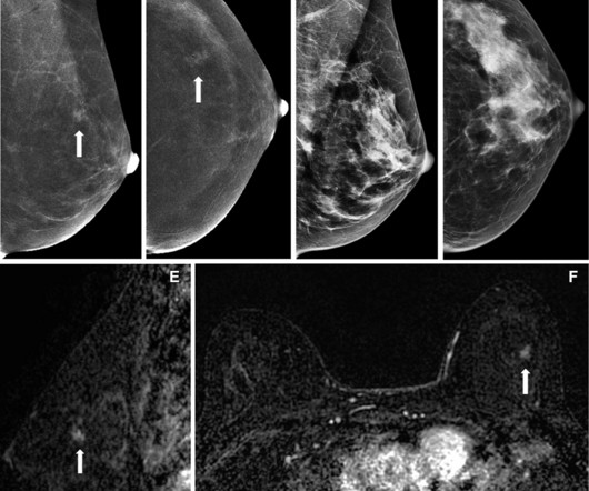
(A) A craniocaudal two-dimensional synthetic mammogram of the right breast shows an irregular mass in the medial breast with pleomorphic calcifications extending posteriorly. (B) B) A craniocaudal magnified mammogram more clearly shows the irregular mass and pleomorphic calcifications in the medial breast. (C)
A team led by Julie Hamzah, MBBS, from Singapore General Hospital, found that symptomatic first breast cancers, dense breasts, and the presence of trabecular thickening on mammography are tied to mammogram detection failure of ipsilateral second breast cancers.
Why would you want to read plain films or thyroid ultrasounds if there are screening mammograms or negative headache brain MRIs ripe for the taking? If you are being paid extra to produce more numbers then why would you want to talk to a clinician on the phone if you could have read another scan during the same amount of time?
A team led by Joao Horvat, MD, from the Memorial Sloan Kettering Cancer Center in New York found that CEM depicted 90% of breast cancers compared with 10% on low-energy mammograms alone and 50% on low-energymammogramswith whole-breast ultrasound. Horvat and co-authors investigated whether the same trend goes for CEM.
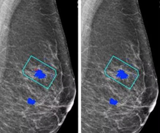
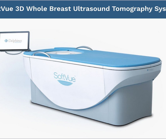
mtaschetta-millane Tue, 07/02/2024 - 09:50 July 2, 2024 — Delphinus Medical Technologies , a pioneering medical imaging company that developed the SoftVue Breast Ultrasound Tomography (UST), announced today the publication of a study comparing mammography in conjunction with SoftVue UST vs mammography alone in women with dense breasts.
House Bill 2411 was introduced in the state by Representative David Cook (R-Globe) and includes eliminating costs for patients for MRI, ultrasound, and diagnostic mammograms. However, out-of-pocket costs for patients can range from $234 for a diagnostic mammogram to more than $1,000 for a breast MRI, according to the organization.
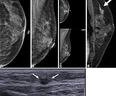
(B) Axial subtracted contrast-enhanced fat-suppressed T1-weighted image from a subsequent-round abbreviated MRI examination performed two years later shows a new 5-mm enhancing mass in the upper outer right breast (arrow), which was not seen on a mammogram performed five months prior. The exam was assessed as BI-RADS category 5.
milla1cf Fri, 07/28/2023 - 23:31 July 28, 2023 — Findings from an accepted manuscript published in the American Journal of Roentgenology (AJR) suggest that for patients with dense breasts undergoing screening in the incidence setting, a commercial AI tool did not provide additional benefit to mammography with supplementary ultrasound (US).
Researchers led by Derek Nguyen, MD, from Duke University in Durham, NC, found that for architectural distortions found by DBT alone with no ultrasound correlate, the malignancy rate was 0% for distortions without atypia versus 20% for distortions with atypia on core needle biopsy. No ultrasound correlate was identified (not shown).
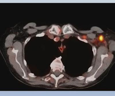
Read more on AuntMinnie.com Related Reading: PET tracer uptake related to COVID-19 vaccines detected in lymphoma patients F-18 FDG lymph node uptake assessed in vaccinated COVID-19 patients Women benefit from scheduling mammograms around COVID vaccine Ultrasound finds swollen glands on patients after COVID-19 vaccine
Gordon pioneered breast ultrasound in Vancouver, BC, and demonstrated its role in detecting cancers missed by mammograms, the SBI said. This award recognizes individuals who have made exceptional strides in advancing breast imaging and have shown extraordinary service to the medical community, the SBI said.
The American Cancer Society recommends starting annual mammogram screenings at age 40. Mammogram Screening Mammogram: Screening mammograms take 2 or more images of each breast. Diagnostic Mammogram: Diagnostic Mammograms are ordered for a variety of reasons and may assess one or both breasts.
Screening breast ultrasounds, in addition to mammograms, save lives. Beginning today, all U.S. women will receive standardized information regarding their breast density. If you have dense breast tissue, be aware and be active.
From each patient, the model extracted 4,096 features from mammograms and selected six related features according to the relationship between tumor size determination and lymph node metastasis. The image is available for use under a Creative Commons license: CC BY-NC-ND 4.0 DEED Attribution-NonCommercial-NoDerivs 4.0 International.
What’s the difference between Screening and Diagnostic Mammogram? During a diagnostic mammogram, the images are analyzed in real-time. During a diagnostic mammogram, the images are analyzed in real-time. Sometimes additional mammogram images are taken.
When 53-year-old photographer and single mom Pia Navales went to the Berkeley Outpatient Center for her annual mammogram in December 2021, she had no reason to suspect any problems. The mammogram identified three masses on her left breast.
That changed in 2023, when Angie underwent a mammogram and breast ultrasound at a Midstate Radiology Associates location offering CARE. Based on Angie’s risk score, she qualified for a breast MRI, a diagnostic tool capable of detecting cancers that mammograms and ultrasounds might miss.
This breast surgery is often performed long before a woman needs her first mammogram , which means that the topic of breast implants’ effects on necessary screenings is rarely discussed before breast augmentation is performed. How often do I need a mammogram? Mammograms should be scheduled every 1 to 2 years until at least age 75.
Topics include the following: Lunit's AI model combines new and existing AI algorithms to filter normal chest radiographs from the radiology workload An exploration of the accuracy and robustness of Lunit's Insight MMG compared with radiologist readers Lunit's AI model tracks mammographic parenchymal patterns as predictive markers for breast cancer (..)
Why Breast Density Matters in Cancer Screening Dense breast tissue affects screening in two key ways: Reduced Visibility : Dense tissue appears white on mammograms, as do tumors, making it harder to detect abnormalities. Inter-radiologist Variation : Assessments can vary up to 33% 1 when different radiologists interpret the same mammograms.
Maybe you have recently moved, changed doctors, or have new insurance and now, are going to a different imaging center for your annual mammogram. Either way, the new imaging center will very often request that you provide the images from your previous mammograms. In this case, the radiologist may recommend a diagnostic mammogram.
Capitol Imaging Services has served tens of thousands of women with our women’s health examinations in screening mammography, diagnostic mammography, ultrasound, bone density studies, breast MRI and breast biopsy. For screening mammograms, we ask for about 20 minutes of a woman’s time. Convenient.
Researchers have developed a wearable ultrasound device that can detect tumors in their early stages and could particularly benefit patients at a high risk of developing breast cancer between routine mammograms.
Ultrasound rounds out the radiologist’s toolkit for supplemental imaging of women with dense breasts. Both handheld and automated ultrasound methods are shown to be effective in detecting mammographically occult cancer in women with dense breast tissue. Advancements in technology are continuously raising the bar for early detection.
We organize all of the trending information in your field so you don't have to. Join 5,000 users and stay up to date on the latest articles your peers are reading.
You know about us, now we want to get to know you!
Let's personalize your content
Let's get even more personalized
We recognize your account from another site in our network, please click 'Send Email' below to continue with verifying your account and setting a password.


Let's personalize your content