This site uses cookies to improve your experience. To help us insure we adhere to various privacy regulations, please select your country/region of residence. If you do not select a country, we will assume you are from the United States. Select your Cookie Settings or view our Privacy Policy and Terms of Use.
Cookie Settings
Cookies and similar technologies are used on this website for proper function of the website, for tracking performance analytics and for marketing purposes. We and some of our third-party providers may use cookie data for various purposes. Please review the cookie settings below and choose your preference.
Used for the proper function of the website
Used for monitoring website traffic and interactions
Cookie Settings
Cookies and similar technologies are used on this website for proper function of the website, for tracking performance analytics and for marketing purposes. We and some of our third-party providers may use cookie data for various purposes. Please review the cookie settings below and choose your preference.
Strictly Necessary: Used for the proper function of the website
Performance/Analytics: Used for monitoring website traffic and interactions
While services for breast and lung cancer screening were temporarily halted, imagers in x-ray, lung ultrasound, and PET/CT were busy examining patients who presented with COVID-19. Her team employed a tracking mechanism for patients who were due for their mammograms once screening operations resumed.
Breast density can often obscure lesions on conventional x-ray mammography, and so other screening modalities such as MRI or ultrasound are often recommended for follow-up. CEM is faster and less costly than MRI and can often be used as a follow-up to an abnormal screening mammogram when it is clinically appropriate.
Using the high-volume single-view chest x-ray professional component (71045-26) as an example, here is the potential impact in Mississippi that was averted by the CAA 24: Using Final CF Locality Reimbursement Change from 2023 Change due to GPCI 2023 $ 8.35 - - 2024 with 1.00 resulting in even lower payments. wGPCI $ 7.64 -8.5% -3.9%
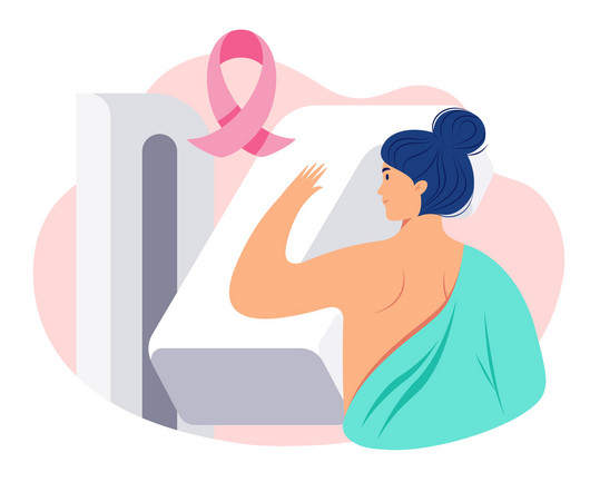
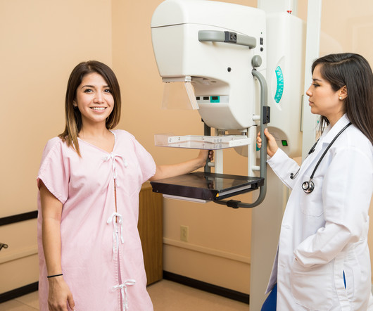
Amidst the battle against this disease, screening mammograms emerge as a crucial tool in early detection and effective treatment. In this blog, we delve into the significance of screening mammograms, their procedure, their benefits, and why they are essential for women’s health. What is a Screening Mammogram?
The studies include two oral presentations and five posters, and address AI advancements for chest x-ray reporting and breast cancer risk assessment, the company said. Lunit plans to highlight seven AI-based studies at the upcoming RSNA meeting.
The American Cancer Society recommends starting annual mammogram screenings at age 40. Mammogram Screening Mammogram: Screening mammograms take 2 or more images of each breast. Diagnostic Mammogram: Diagnostic Mammograms are ordered for a variety of reasons and may assess one or both breasts.
With three outpatient imaging locations throughout Boise, Meridian, and Eagle, Intermountain Medical Imaging offers diagnostic imaging procedures in a calm and comforting environment, including MRI, CT, Ultrasound, X-ray, Interventional Radiology, and Mammography. “St. This new partnership between St.
milla1cf Tue, 08/29/2023 - 15:55 August 29, 2023 — Mammograms are an essential part of preventive healthcare, and when an initial review reveals a suspicious lesion, additional imaging and/or invasive breast biopsies could be the next step in diagnosis. to adopt the recently available Imagio Breast Imaging System.
At PURE Mammography, we offer 3D breast mammography as well as ultrasound imaging for breasts and other areas of the body. In addition to mammograms and ultrasounds, women should know about the various other breast screenings that are performed. Breast Ultrasound A breast ultrasound is different than an x-ray.
Have you ever jumped out of bed, excited for your mammogram appointment? At Clermont Radiology’s Women’s Center, we offer Digital Mammography, Ultrasound, and DEXA Scans. A mammogram is a low dose x-ray of the breast. Our Women's Center performs both screening and diagnostic mammograms. Probably not!
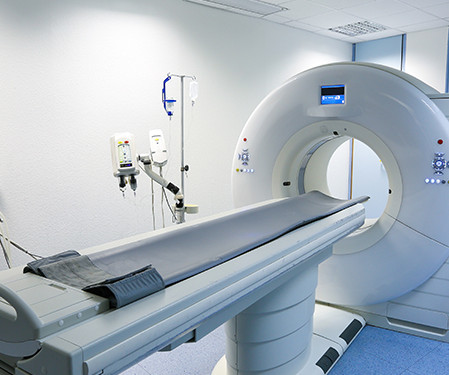
There are several types of imaging tests that physicians use to detect cancer in patients: X-Ray, Computed Tomography (CT), Magnetic Resonance Imaging (MRI), Ultrasound (US), Nuclear Medicine, and Positron Emission Tomography (PET). It is also used to determine the progression of existing cancerous masses.
Try as we might, good health practices tend to be neglected, especially regarding preventive health screenings like mammograms. Research shows that one in four women who should be getting regular mammograms don’t. Women with an average risk for breast cancer should begin mammogram screening every year starting at 40.
The product can be used to visualize known or suspected lesions of the breast in adults, as an adjunct to mammography and/or ultrasound. Through the use of iodine-based x-ray contrast agents, CEM can allow for better visualization of abnormalities in breast tissue that may not be visible with standard mammography.3
For some patients, tissues can overlap potentially hiding signs of breast cancer; in other cases, the tissue and cancer can show up as white on a mammogram making diagnosis more difficult. Combined screening with mammography and ultrasound in a population-based screening program. Breast Cancer Facts and Statistics. Eur J Radiol.
Medical Imaging of Fredericksburg’s services include X-Ray, CT, PET/CT, MRI, 3D mammograms, Ultrasound and a variety of other health scans. Patients have given an over 95% satisfaction score – a reflection of the level of commitment the physicians and staff have to the community they serve.
With an X-ray, Ultrasound, Mammogram and CT scan at our disposal, we needed to have a radiologist who could read all these modalities, and give us results in the shortest time possible to enable us to give the best medical care possible” she says. MHRG devised a 24/7 remote radiology solution for the Clinic.
All of the annual scheduled services such as mammograms can now be scheduled, as well as imaging prescribed by physicians for the care of their patients. Patients may also schedule mammograms directly at our facilities in these same locations. Christopher Newman, Chief Medical Officer of Mary Washington Healthcare.
A trained onsite sonologist with remote interpretation of the ultrasound studies helped them to fulfil their vision. Lives MHRG have touched: “A self-referred 51-year-old with a cough who requested a chest x-ray and a mammogram to be diagnosed with metastatic breast cancer having been treated for pneumonia over and over again…. “45-year-old
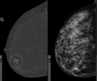
Women with dense breasts are BOTH more likely to develop breast cancer and more likely to have that cancer missed on a mammogram [5] Fig. 1 – Cancer on a mammogram of a fatty vs a dense breast What is Dense Breast Tissue? Breast density is determined through a mammogram and described as one of four categories (Fig.
Mammography is an X-Ray of the breasts carried out on a machine dedicated to it because the breast is not of uniform thickness and requires a dedicated unit. Hence, mammograms carried out anywhere can now be viewed by expert breast radiologists in any part of the world thanks to teleradiology. Age 40-50 Once in 2 years 3.
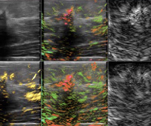
Our radiologists with breast imaging expertise can re-evaluate mammograms, breast MRIs, and ultrasounds. Ultrasound Obstetric and Gynecologic Ultrasounds : Subtle findings such as ovarian cysts or fetal abnormalities often require expert review to confirm a diagnosis.
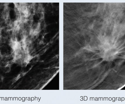
With traditional mammograms, it can be difficult to detect cancers in dense breasts because the images are acquired in two dimensions and the breast is a three-dimensional structure. Usually these overlapping areas can be cleared with additional images and ultrasound but “false positive” call-backs cause a great deal of anxiety for women.
It all started when Wilhelm Conrad Röntgen discovered X-rays in 1895. After working for weeks in his lab experimenting on the production of ‘strange rays’, which he referred to as ‘X’, he asked his wife Anna Bertha to lend ‘a hand’, the left one to be precise, which he used to produce the first X-ray image.
We organize all of the trending information in your field so you don't have to. Join 5,000 users and stay up to date on the latest articles your peers are reading.
You know about us, now we want to get to know you!
Let's personalize your content
Let's get even more personalized
We recognize your account from another site in our network, please click 'Send Email' below to continue with verifying your account and setting a password.


Let's personalize your content