This site uses cookies to improve your experience. To help us insure we adhere to various privacy regulations, please select your country/region of residence. If you do not select a country, we will assume you are from the United States. Select your Cookie Settings or view our Privacy Policy and Terms of Use.
Cookie Settings
Cookies and similar technologies are used on this website for proper function of the website, for tracking performance analytics and for marketing purposes. We and some of our third-party providers may use cookie data for various purposes. Please review the cookie settings below and choose your preference.
Used for the proper function of the website
Used for monitoring website traffic and interactions
Cookie Settings
Cookies and similar technologies are used on this website for proper function of the website, for tracking performance analytics and for marketing purposes. We and some of our third-party providers may use cookie data for various purposes. Please review the cookie settings below and choose your preference.
Strictly Necessary: Used for the proper function of the website
Performance/Analytics: Used for monitoring website traffic and interactions
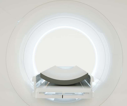
In 2023 compared with 2022, the total volume of positron emission tomography (PET) scans increased 10.2% year over year, according to the newly published IMV 2024 PET Market Summary Report. In 2023, the average number of PETscans per fixed PET site (versus mobile PET) increased 6.7%
Over the same period between the two years, the average number of PETscans performed per installed fixed PET/CT systems increased by 8.2% -- a finding that indicates that much of the increased procedure volumes have been absorbed by existing PET system capacity. Davin Korstjens of IMV Medical Information Division.
The way physicians identify illness is changing due to advances in medicalimaging, which make early diagnosis quicker, more precise, and less invasive. These technologies, ranging from high-resolution MRIs to state-of-the-art CT scans, can give doctors the ability to spot possible health problems before symptoms even show up.
Centers for Medicare and Medicaid Services (CMS) announced on October 13 that it has lifted its coverage limit of one beta-amyloid PETscan per lifetime for patients with Alzheimer’s disease. Medicare coverage decisions for amyloid PETscans will now be made by local Medicare Administrative Contractors (MACs).
In brief, the deep learning-based AI model (called “JuST_BrainPET”) is designed to automatically segment metabolic tumor volume (MTV) from surrounding healthy tissue on brain PETimaging, a key step in medicalimaging analysis, the researchers explained. Image courtesy of the Journal of Nuclear Medicine.
The technology could allow clinicians to detect multiple biomarkers at once and improve the spatial resolution of brain imaging for applications in cancer as well as neurodegenerative disease, according to principal investigator Lars Furenlid, PhD, a professor of medicalimaging at the University of Arizona.
The MedicalImaging and Technology Alliance (MITA) has raised concerns ove. Read more on AuntMinnie.com Related Reading: ACR urges full coverage for amyloid PETscans CMS proposal covers PETscans for Alzheimer’s disease MITA praises FDA for Leqembi approval FIND Act reintroduced in U.S.
PETscans typically used for diagnosing prostate cancer may have value in detecting inflammatory bowel disease (IBD), according to a case series published December 8 in Clinical and Experimental Gastroenterology.
Increased use of medicalimaging by other specialties (turf battles) HOTTEST CLINICAL PROCEDURE 1. Hybrid CT/PETscanning 2. Introduction of 16-slice CT scanners 2. Ongoing shortage of qualified staff BIGGEST THREAT TO RADIOLOGY 1. Ongoing shortage of qualified staff 2. 165-175).
"Radiologists are uniquely positioned to educate other healthcare providers on how to properly remove personal health information from radiologic imaging files," wrote Stern and colleague William Weadock, MD, also of the university. PowerPoint presentations that include medicalimages are a valuable educational tool, Stern and Weadock noted.
TRA MedicalImaging is proud to offer PSMA PET, a cutting-edge imaging method that enables us to detect prostate cancer with greater precision than conventional imaging techniques. Our compassionate team is here to support you every step of the way, close to home, with the latest in imaging technology.
When your doctor orders an imaging exam, one of the first questions you might have is: How much will this cost? At TRA MedicalImaging, we believe in transparent pricing and helping you navigate the complexities of insurance coverage, out-of-pocket costs, and financial assistance options. Why Choose TRA MedicalImaging?
Medicalimaging has evolved over centuries, starting with X-rays in 1895, progressing to CT, MRI, and PETscans. The post Revolutionising Healthcare: A Historical Perspective on MedicalImaging appeared first on Open Medscience.
Teleradiology & Radiology data for artificial intelligence (AI) Introduction: Embark on a journey into the world of medicalimaging as we unravel the distinctions between two powerful diagnostic tools—Computed Tomography (CT) scans and Positron Emission Tomography (PET) scans.
State-of-the-Art Technology Not all imaging centers offer the same equipment, and newer technology often leads to more precise imaging and quicker diagnoses. Look for centers that offer advanced technologies like 3D mammography, PSMA PETscans for prostate cancer, and low-dose CT scans for lung cancer screening.
PETscans are crucial for detecting metabolic activity, providing valuable insights into cancer, neurological disorders, and cardiovascular diseases. The post How to Read a PETScan: A Basic Understanding appeared first on Open MedScience.
Medicalimaging crucially enhances oncology, aiding early cancer detection and effective treatment planning. The post The Transformative Impact of MedicalImaging in Oncology appeared first on Open Medscience.
This description highlights the significant advancements in medicalimaging that are transforming healthcare. Advanced algorithms can analyze images more quickly and accurately, aiding in early diagnosis and treatment planning. This aids in the early detection and monitoring of diseases.
Advances in medicalimaging technology have significantly improved diagnostic accuracy, enabling earlier detection and more personalised treatments. The post Advances in MedicalImaging Technology: Unlocking the Mysteries of the Human Body appeared first on Open MedScience.
When your doctor orders an imaging exam, one of the first questions you might have is: How much will this cost? At TRA MedicalImaging, we believe in transparent pricing and helping you navigate the complexities of insurance coverage, out-of-pocket costs, and financial assistance options. Why Choose TRA MedicalImaging?
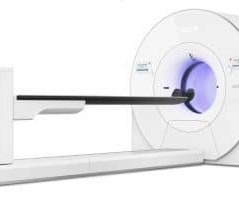
Additionally, Omni Legend offers a streamlined, simple solution that helps enable technologists to increase efficiency, enhance patient care, and reduce potential radiation exposure to medical staff vii.” iii] Based on orders data of GE HealthCare PET/CT systems since 2010. [iv] Eur J of Nucl Med Mol Imaging 49, 3740–3749 (2022).
TRA MedicalImaging is proud to offer PSMA PET, a cutting-edge imaging method that enables us to detect prostate cancer with greater precision than conventional imaging techniques. Our compassionate team is here to support you every step of the way, close to home, with the latest in imaging technology.
The authors discuss several AI strategies for improving tumor detection, including a holistic approach that integrates data from various imaging techniques such as MRI, CT scans, and PETscans, along with genomic information and patient histories.
MRI technologies are also particularly beneficial for scanning the brain, spine, soft tissues in the joints, and the interior structures of bones. The post Magnetic Resonance Imaging (MRI): A Leading Imaging Modality Because of its Diagnostic Versatility appeared first on Associates in MedicalImaging.
PACS – Picture Archiving and Communication System; a system involved in acquiring the medicalimages, transmission, viewing, storage, and retrieval of same images. of imaging modalities, storage space, no. Basically, PACS is an electronic version of the file room and reading room for radiologists.
EXPLORER, the world’s first medicalimaging scanner to produce a 3-D picture of the whole human body. The post EXPLORER PET-CT scanner: a total body experience appeared first on Open Medscience.
In all cases, your doctor may recommend multiple forms tests if you display symptoms and have a history of asbestos exposure, including bloodwork and imaging. Imaging for Mesothelioma Medicalimaging serves several purposes. It assists with diagnosing the presence of tumors and determining if the cancer has spread.
DICOM Image Format is an international standard to transmit, store, retrieve, print, process, and display medicalimaging information. DICOM allows transmitting medicalimaging data to devices like scanners, servers, workstations, printers, network hardware, and PACS.
How Much Does a PET/CT cost? A lot can go through your mind when your physician orders a PET/CT for you. Along with the normal anxiety associated with needing a medicalimaging study, worrying about your out-of-pocket expense is a reasonable concern. But, first, let’s understand what a PET/CT is and why it is used.
Diagnostic imaging in motor neurone disease (MND) is crucial for early detection, disease monitoring, and differentiating from other conditions. The post Motor Neurone Disease: Diagnosis and Future Research Insights appeared first on Open MedScience.
Positron Emission Tomography Imaging has advanced with cutting-edge technologies, enhancing diagnostic accuracy and expanding clinical applications dramatically. The post Breakthroughs in Positron Emission Tomography Imaging appeared first on Open MedScience.
The following is the list of candidates for the 2024 edition of the Minnies, AuntMinnie.com 's campaign to recognize the best and brightest in medicalimaging. Image from Gregory Barnes, MD, PhD, of the Norton Children's Autism Center, et al. Image from Terril Verplaetse, PhD, of Yale University, et al.
To analyse the CT scan data, the researchers used a technique called radiomics, which can extract information about the patient’s disease from medicalimages that can’t be easily seen by the human eye. size, location, spiculation status), but also includes PETscan findings.
We organize all of the trending information in your field so you don't have to. Join 5,000 users and stay up to date on the latest articles your peers are reading.
You know about us, now we want to get to know you!
Let's personalize your content
Let's get even more personalized
We recognize your account from another site in our network, please click 'Send Email' below to continue with verifying your account and setting a password.


Let's personalize your content