This site uses cookies to improve your experience. To help us insure we adhere to various privacy regulations, please select your country/region of residence. If you do not select a country, we will assume you are from the United States. Select your Cookie Settings or view our Privacy Policy and Terms of Use.
Cookie Settings
Cookies and similar technologies are used on this website for proper function of the website, for tracking performance analytics and for marketing purposes. We and some of our third-party providers may use cookie data for various purposes. Please review the cookie settings below and choose your preference.
Used for the proper function of the website
Used for monitoring website traffic and interactions
Cookie Settings
Cookies and similar technologies are used on this website for proper function of the website, for tracking performance analytics and for marketing purposes. We and some of our third-party providers may use cookie data for various purposes. Please review the cookie settings below and choose your preference.
Strictly Necessary: Used for the proper function of the website
Performance/Analytics: Used for monitoring website traffic and interactions
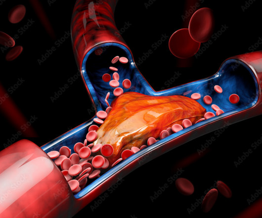
While anticoagulant medications to prevent further clot formation is a primary treatment in nonemergency cases, the use endovascular procedures such as arterial and venous thrombectomy to treat more severe cases has become increasingly common, according to McNamara.
Radiologists are medical doctors who specialize in interpreting imaging studies like X-rays, CT scans, MRIs, and ultrasounds to diagnose and guide treatment for various conditions. This rigorous training covers all imaging modalities, from X-rays to advanced techniques like MRI and PET/CT scans.
Introduction: Within the realm of medical imaging, the world of X-rays unveils a captivating interplay between science and artistry. This blog delves into the fascinating world of X-ray perspectives, exploring how the science behind medical imaging meets the artistry that brings these images to life.
Teleradiology & Radiology data for artificial intelligence (AI) Introduction: “Illuminating Shadows” invites you on a comprehensive journey into the fascinating world of X-ray imaging. Chapter 1: Introduction to X-ray Imaging An overview of the importance of X-ray imaging in healthcare.
Benefits of Teleradiology to Telehealth Introduction: “Behind the Beams” is an intriguing journey that delves into the intricate world of X-ray technology, uncovering the perfect fusion of art and science that powers this essential diagnostic tool. How each modality serves specific clinical needs and diagnostic challenges.
Closeup of X-ray photography of human brain Introduction: In the world of modern medicine, there exists a fascinating blend of art and science, where the careful use of technology and technique converges to reveal the hidden truths within the human body.
Also featured is the next-generation Image Guided Therapy System – Azurion 7 B20/15 biplane configuration, providing superb positioning capability for easier patient access during minimallyinvasiveprocedures, faster system movement, and full table side control of all components.
Designed to improve productivity and help care teams make the right decisions faster, treat more patients, and achieve better outcomes, the new interventional system features enhanced 2D and 3D imaging and X-ray detector positioning flexibility.
As technology evolved, doctors turned to imaging-guided procedures using computed tomography (CT) or X-ray technology. Considered a minimallyinvasiveprocedure, image-guided epidural injections for back pain may be administered at a doctor’s office, surgical center or hospital imaging center.
Mammography Mammography is a specialized imaging service that uses low-dose X-rays to examine breast tissue. Let’s explore these specialized imaging services and understand their significance in women’s health. Prevention: Early detection and treatment can prevent fractures and maintain a higher quality of life.
AP Chest X-ray demonstrating placement of 3 MitraClips over the tricuspid valve. A: AP chest X-ray demonstrating placement of 3 MitraClips over the tricuspid valve (yellow arrow) B: Coronal chest CT showing 3 MitraClips over the tricuspid valve in the right atrioventricular septum (red arrow). Name the cardiac device.
As demand for minimallyinvasiveprocedures continues to grow, GE HealthCare is committed to helping clinicians use image guidance technologies to their full potential by removing barriers with the goal of helping providers achieve better clinical and operational outcomes.
We organize all of the trending information in your field so you don't have to. Join 5,000 users and stay up to date on the latest articles your peers are reading.
You know about us, now we want to get to know you!
Let's personalize your content
Let's get even more personalized
We recognize your account from another site in our network, please click 'Send Email' below to continue with verifying your account and setting a password.


Let's personalize your content