This site uses cookies to improve your experience. To help us insure we adhere to various privacy regulations, please select your country/region of residence. If you do not select a country, we will assume you are from the United States. Select your Cookie Settings or view our Privacy Policy and Terms of Use.
Cookie Settings
Cookies and similar technologies are used on this website for proper function of the website, for tracking performance analytics and for marketing purposes. We and some of our third-party providers may use cookie data for various purposes. Please review the cookie settings below and choose your preference.
Used for the proper function of the website
Used for monitoring website traffic and interactions
Cookie Settings
Cookies and similar technologies are used on this website for proper function of the website, for tracking performance analytics and for marketing purposes. We and some of our third-party providers may use cookie data for various purposes. Please review the cookie settings below and choose your preference.
Strictly Necessary: Used for the proper function of the website
Performance/Analytics: Used for monitoring website traffic and interactions
Images from the world's largest radiology conference include new technologies and the latest advances in MRI, CT, nuclear medicine, X-ray, artificial intelligence, and PACS/enterprise imaging.
Radiologists, technologists, administrators, and industry professionals can find information and conduct e-commerce in MRI, mammography, ultrasound, x-ray, CT, nuclear medicine, PACS, and other imaging disciplines.
Radiologists, technologists, administrators, and industry professionals can find information and conduct e-commerce in MRI, mammography, ultrasound, x-ray, CT, nuclear medicine, PACS, and other imaging disciplines.
Clinical use of imaging equipment accounts for more than 50% of greenhouse gas emissions in the radiology department, with MRI and CT equipment major contributors, researchers have reported. Of these emissions, MRI was responsible for 48% (2.2 Use of PACS made up 11% of departmental greenhouse gas emissions (0.48 kt CO 2 e).
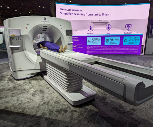
CHICAGO -- GE HealthCare (GEHC) brought new CT and MRI scanners and a range of AI software applications to McCormick Place for RSNA 2023. The scanner also comes with True Enhance DL, an AI-based application that generates deep learning-based monochromatic-like images from a single-energy x-ray acquisition.
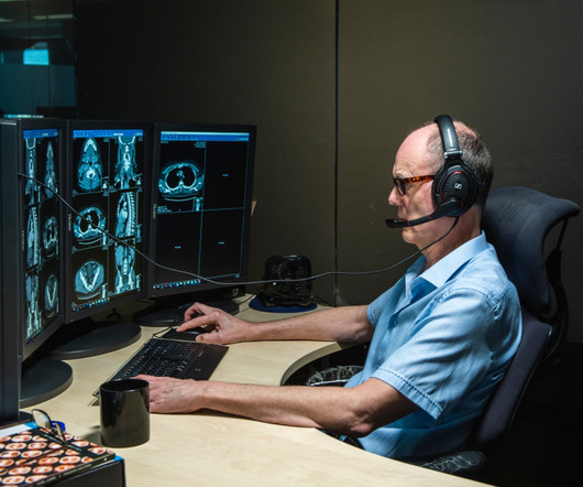
All this is possible because of PACS. PACS – Picture Archiving and Communication System; a system involved in acquiring the medical images, transmission, viewing, storage, and retrieval of same images. Basically, PACS is an electronic version of the file room and reading room for radiologists.
The new innovations Philips announced at #RSNA23 included next generation ultrasound systems that increase diagnostic confidence and workflow efficiency, the world’s first and only mobile MRI system with helium free operations, and new AI-enabled cloud solutions that enhance radiology efficiency and clinical confidence.
Angiography imaging systems have the second highest energetic impact behind MRI scanners, according to the researchers. To address this gap in knowledge, the researchers collected data from RIS log files and PACS reports from a biplane angiography system (Azurion 7 B20, Philips ) over a six-weeks period.
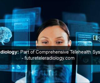
Explore how technologies like digital X-ray, CT, and MRI have paved the way for efficient image acquisition and transmission in teleradiology. Picture Archiving and Communication Systems (PACS): Highlight the significance of PACS in teleradiology.
Diagnosis often depends on the interpretation of tests like of X-Rays, CT Scan, MRI, PET CT Scan , etc. They should have PACS software as well. PACS is an electronic platform where radiology images that connected with medical automation systems. They should have PACS software as well.
Inception and Exploration: Revealing the Genesis Explore the formative days of radiology, ignited by Wilhelm Roentgen’s groundbreaking discovery of X-rays in 1895. Discuss how PACS has redefined collaboration and accessibility within the dynamic realm of radiology workflows.
Explore how systems are designed to handle data from modalities such as X-ray, CT, MRI, and ultrasound, providing a comprehensive diagnostic overview. Unified Picture Archiving and Communication Systems (PACS): Highlight the importance of unified PACS in teleradiology integration.
Today’s teleradiology is integrated with the latest PACS, AI enabled workflow, internet connectivity with wide bandwidth and an experienced back-end support team that has made the reporting possible with short TAT. The ultrasound machine and mobile X-ray machines are proving it with their wire-less and cloud-computing features.
Applications of Remote Reporting: Explore the diverse applications of remote reporting across various medical imaging modalities (X-ray, CT, MRI, etc.). Explore the interoperability of teleradiology systems with hospital information systems (HIS) and picture archiving and communication systems (PACS).
DICOM allows transmitting medical imaging data to devices like scanners, servers, workstations, printers, network hardware, and PACS. In this blog article, we will explain the fundamentals of DICOM technology, its impact on clinical practice and radiology workflows, and dig deeper into enhanced functionality offered by modern PACS systems.
Hub Prior to the pandemic, TMC were aware of a growing need for acute reporting services ranging from neuro MRI ad hoc reporting to Emergency CT daytime cover to sub-specialist short turn around reporting. And new to TMC’s repertoire is a novel service, bringing AI to its clients without them knowing it.
tesla MRI AI body composition analysis Cardiac PET Cryo/thermoablation CT colonography Genicular artery embolization Hyperpolarized xenon-129 MRI PET/MRI Photon-counting CT Radiomics Theranostics Whole-body MRI screening Image of the Year 3D PET/MR image. tesla brain MRI scans. tesla brain MRI scans.
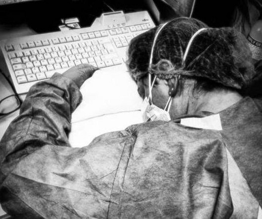
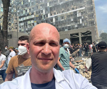
X-ray and ultrasound machines were badly damaged in a rocket attack on Ukraine's largest children's hospital on July 8, according to radiologist Stanislav Rebenkov, MD. In particular, the facility lacks a modern PACS and clinical decision-support tools. It will take months to recover, he said.
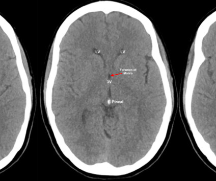
The goal of this multi-part blog is as follows: To cover the basics of how to look at a CT brain and quickly identify life threat Review the literature supporting the major ED indications Discuss special considerations, such as when to use contrast, angiography, or MRI instead. X-ray attenuation. Shaw AS, Prokop M. McKetty MH.
Healthcare examples : Chatbots for billing and scheduling or filtering and organizing data within a medical device, such as an MRI or CT scanner. These AI tools act as intelligent assistants, sifting through massive amounts of scans, like X-rays, CTs and MRIs, flagging potentially critical abnormalities.
It all started when Wilhelm Conrad Röntgen discovered X-rays in 1895. After working for weeks in his lab experimenting on the production of ‘strange rays’, which he referred to as ‘X’, he asked his wife Anna Bertha to lend ‘a hand’, the left one to be precise, which he used to produce the first X-ray image.
We organize all of the trending information in your field so you don't have to. Join 5,000 users and stay up to date on the latest articles your peers are reading.
You know about us, now we want to get to know you!
Let's personalize your content
Let's get even more personalized
We recognize your account from another site in our network, please click 'Send Email' below to continue with verifying your account and setting a password.


Let's personalize your content