This site uses cookies to improve your experience. To help us insure we adhere to various privacy regulations, please select your country/region of residence. If you do not select a country, we will assume you are from the United States. Select your Cookie Settings or view our Privacy Policy and Terms of Use.
Cookie Settings
Cookies and similar technologies are used on this website for proper function of the website, for tracking performance analytics and for marketing purposes. We and some of our third-party providers may use cookie data for various purposes. Please review the cookie settings below and choose your preference.
Used for the proper function of the website
Used for monitoring website traffic and interactions
Cookie Settings
Cookies and similar technologies are used on this website for proper function of the website, for tracking performance analytics and for marketing purposes. We and some of our third-party providers may use cookie data for various purposes. Please review the cookie settings below and choose your preference.
Strictly Necessary: Used for the proper function of the website
Performance/Analytics: Used for monitoring website traffic and interactions
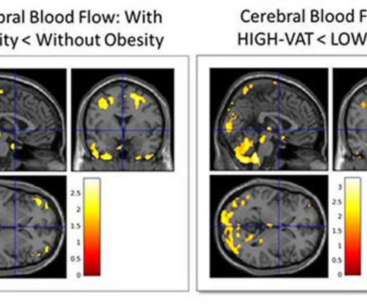
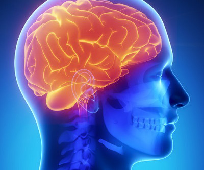
CHICAGO -- Characterizing an individual's type of body fat using body MRI can help predict Alzheimer's disease risk up to 20 years before symptoms manifest, according to research results presented December 2 at the RSNA meeting.
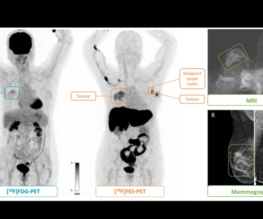
Between December 2018 and January 2021, the group enrolled 41 female participants (median age, 56 years) who underwent both PETscans. F-18 FES-PETscan shows a second primary tumor (grade 1, lobular) in the left breast and malignant lymph nodes (T2N1M0) that are not visible on the F-18 FDG-PETscan.
A PET radiotracer for diagnosing Alzheimer’s disease may also be used to measure vascular brain changes in patients during PET/MRIscans, according to a study published December 7 in the Journal of Nuclear Medicine. The researchers enrolled 20 participants. Image courtesy of the Journal of Nuclear Medicine.
MRI: Uses magnets and radio waves to create detailed images of tissues and organs. CT Scans: Provides cross-sectional images of the body for a more detailed view. PETScans : Help detect diseases at a cellular level, often used for cancer detection and brain disorders.
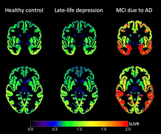
A PET/MRI study has provided insights into the neurobiology of late-life depression, with researchers reporting that tau protein – a key protein involved in Alzheimer’s disease – does not appear to be involved in the condition. However, further studies are needed to determine if tau PETscans could help play a role, they noted.
Read more on AuntMinnie.com Related Reading: FDA grants full approval to Alzheimer's disease drug Leqembi CMS rethinks limit on PETscans for Alzheimer’s disease patients FDA doubles MRIscans needed for Aduhelm patients CMS to limit coverage of new drugs for Alzheimer's disease CMS scrubs rule restricting PET tracer coverage
This persistent inflammation increases atrophy of gray matter in the brain, yet is difficult to assess on MRIscans, the authors explained. The researchers performed F-18 PBR06 PETscans on 22 patients with MS and eight healthy controls. Image courtesy of Clinical Nuclear Medicine.
MRI is the imaging gold standard for diagnosis, yet identifying the disease using this method remains challenging, the researchers wrote. In all three cases, gadolinium-enhanced MRIscans did not show abnormalities. F-18 FDG-PET and MRIscans of three patients. (A.1-B.1)
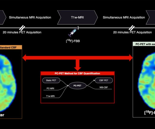
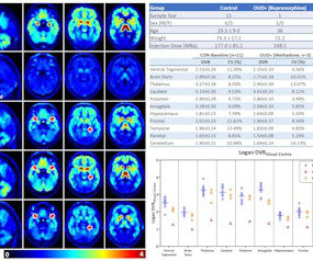
All subjects underwent 90 minute dynamic whole-body PETscans using a dedicated research scanner called PennPET Explorer. Prior to PETscanning, subjects also underwent T1-weighted MRI brain imaging to create templates to segment the PET brain data into 10 regions of interest.
According to clinical trials, amyloid PETscans showed that donanemab reduced amyloid plaques by up to 84% after 18 months of treatment. Safety warnings include that the drug can cause amyloid-related imaging abnormalities, which are detected by MRI and present as temporary swelling in an area or areas of the brain.
tesla MRI AI body composition analysis Cardiac PET Cryo/thermoablation CT colonography Genicular artery embolization Hyperpolarized xenon-129 MRIPET/MRI Photon-counting CT Radiomics Theranostics Whole-body MRI screening Image of the Year 3D PET/MR image. tesla brain MRIscans.
Microglia are immune cells in the brain that are thought to have a role in MS disease progression but cannot be seen by a routine MRI. The team developed a technique called F18 PBR 06 PET imaging. The newly published study involved performing PETscans on 22 people with MS and eight healthy controls.
In this case, a 43-year-old man underwent an MRIscan that showed no contrast enhancement, yet hyperintensities were apparent in the patient’s left thalamus and frontoparietal region. Thus, the clinicians performed an additional PETscan with an amino acid radiotracer (F-18 FET) for further diagnosis of a suspected glioma.
A group in England has established PET imaging as a new approach for studying gait – an excellent indicator of physical, emotional, and mental health, according to a study published February 6 in NeuroImage. F-18 FDG-PETscans allow clinicians to measure the brain's energy demands based on glucose metabolism.
The team investigated any associations between brain MRI volumes -- as well as amyloid and tau uptake on PETscans -- with body mass index (BMI), obesity, insulin resistance, and abdominal fatty tissue in cognitively healthy midlife individuals. The green colors are the normal white matter. Image courtesy of the RSNA.
All patients underwent F-18 PI-2620 PET imaging, as well as amyloid PET, MRI, and neuropsychologic tests at baseline and at follow-up after one year. in 15 patients with negative amyloid PETscans; 1.18 in in 20 late-onset patients, and 1.54 in 17 early-onset patients.
Read more on AuntMinnie.com Related Reading: CMS to cover Alzheimer's drugs for patients enrolled in registry Will lecanemab approval increase PETscan volumes? FDA approves new drug for Alzheimer's disease FDA doubles MRIscans needed for Aduhelm patients MRI sheds light on effects of aducanumab Alzheimer's drug
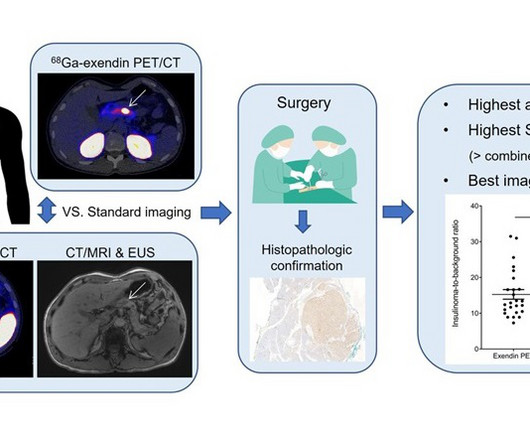
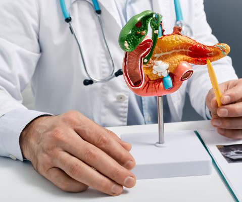
A new PETscan has shown promise in reliably detecting benign tumors in the pancreas that cannot be detected by various imaging techniques such as CT, MRI, and PETscans.
A new PETscan has shown promise in reliably detecting benign tumors in the pancreas that cannot be detected by various imaging techniques such as CT, MRI, and PETscans.
iPET is AI-based software that denoises, sharpens organ boundaries, and improves resolution in PET and SPECT scans. Scans processed with the algorithm can also provide additional details by using an overlay of an MRI or CT image of the same region, the company said.
Read more on AuntMinnie.com Related Reading: Abbreviated MRI protocols effective for emergency applications Can AI help in diagnosing Alzheimer's disease? Will lecanemab approval increase PETscan volumes? Brain PET patterns emerge in early Alzheimer's patients
Read more on AuntMinnie.com Related Reading: CMS to cover Alzheimer's drugs for patients enrolled in registry Will lecanemab approval increase PETscan volumes? FDA approves new drug for Alzheimer's disease CMS delays decision on amyloid PET coverage MRI sheds light on effects of aducanumab Alzheimer's drug
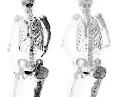
Read more on AuntMinnie.com Related Reading: NaF-PET shows bone formation in psoriatic arthritis patients PET/MRI provides new insights into knee osteoarthritis NaF-PET reveals aortic wall injuries NaF-PETscans reveal plaque -- and possible risk of stroke Can deep learning monitor lesions on F-18 NaF PET/CT?
Still, these studies have included small samples and have not focused on developing an MRI biomarker. LEADS participants receive a standard clinical evaluation and Magnetic Resonance Imaging ( MRI ) scanning as well as amyloid and tau PETscanning and fluid biomarker assessments on an annual basis.
milla1cf Thu, 07/27/2023 - 23:20 July 27, 2023 — Scientists have found a new use for copper in magnetic resonance imaging (MRI) contrast agent design, that could help to create better images which help doctors diagnose patients’ conditions more easily and safely.
The company has been developing human digital twin technology at the organ and lesion level, as well as foundational AI models designed to extract actionable insights from MRI, CT, and PETscans. through the CE and UKCA marks, some of which are 510(k) cleared by the U.S.
Reimbursement barriers Unfortunately, until recently, there were significant barriers to receiving amyloid PET imaging in some regions. Centers for Medicare and Medicaid Services (CMS), for example, did not cover amyloid PETscans outside of clinical trials for Medicare beneficiaries and only covered one scan per patient’s lifetime.
announced a research collaboration agreement with the Alzheimer’s Disease Neuroimaging Initiative 4, ADNI4, on the use of Meilleur’s [F-18]NAV-4694, an investigational imaging agent, in Positron Emission Tomography (PET) scans to assess the status amyloid plaque in the brain.
The answer to many of these questions could be just one—MRI. Offer Versatility and Clarity to Your Doctors and Patients MRI, or Magnetic Resonance Imaging , could be the most versatile imaging technology available to members of the medical community.
Medical imaging has evolved over centuries, starting with X-rays in 1895, progressing to CT, MRI, and PETscans. The post Revolutionising Healthcare: A Historical Perspective on Medical Imaging appeared first on Open Medscience.
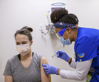
This can look like a clinically significant finding on cancer imaging, including chest CTs, PETscans, mammography, and breast MRI. A small but significant number of people experience swollen lymph nodes as a side effect of receiving a COVID vaccine. … Continue reading →
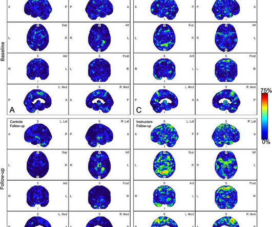
All participants underwent a PETscan of the head to evaluate and quantify amyloid changes. The military instructors filled out a digital log with the number of exposures to explosions, including the firing of weapons. The control participants were evaluated at similar time points.
There’s a strong and innovative imaging department here, which will allow us to use MRI and sophisticated PETscanning to guide treatments, as well as outstanding urology and medical oncology departments, to provide the most comprehensive and multidisciplinary treatment recommendations.”
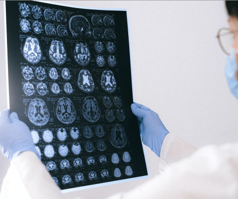
Review Paper on AI and Cancer Detection Professor Pegah Khosravi and her team of researchers explore how artificial intelligence (AI) can enhance anomaly detection in MRIscans to advance precision medicine. By leveraging these methods, a more thorough analysis of MRI data is ensured.
Cutting-Edge Imaging Technologies: PET-CT Scans: Examine how Positron Emission Tomography combined with Computed Tomography (PET-CT) scans offer enhanced imaging, providing information about both structure and metabolic activity.
CT Scan-Guided Biopsy: This technology better identifies the location of the growth, so that a sufficient number of cells can be gathered from the tumor for analysis. PET/CT Scan: A positron emission tomography (PET) scan involves injecting a radioactive, sugar-based substance and observing it accumulate in the potentially cancerous cells.
Long COVID In a study published in Medical Hypotheses , a French group presented a theory regarding the brain fog experienced in long COVID, based on brain patterns identified in patient PETscans. Further prospective longitudinal MRI studies are essential to elucidate causality in this context.
MRI: This procedure offers a detailed look at the body’s soft tissues, including locations of tumors and if malignant mesothelioma has started to spread. PETScan: These scans create 3D images that offer greater insight into whether a thickened pleura or peritoneum is malignant mesothelioma or a benign condition.
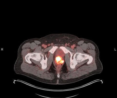
Results demonstrated high detection rates (% positive PETscans) even at low PSA levels. Conventional imaging techniques such as CT and MRI are limited in the information they may provide. The SPOTLIGHT study evaluated POSLUMA in men with suspected prostate cancer recurrence based on elevated PSA following prior therapy.
PACS works as a host that integrates the radiological images acquired from different radiological imaging modalities (X-Ray, Ultrasound, CT, MRI , PETScan, Nuclear Medicine, etc…) with a network of information system (RIS and or HIS), EMR, different work stations and image storage/archival system.
Diagnosis often depends on the interpretation of tests like of X-Rays, CT Scan, MRI, PET CT Scan , etc. It has adhered to the globally standardized reporting format of X-Rays, CT Scan, MRI, PET CT Scan, etc. Diagnosis Radiology images like CT Scan, PETScan, X Rays are needed for diagnosing.
We organize all of the trending information in your field so you don't have to. Join 5,000 users and stay up to date on the latest articles your peers are reading.
You know about us, now we want to get to know you!
Let's personalize your content
Let's get even more personalized
We recognize your account from another site in our network, please click 'Send Email' below to continue with verifying your account and setting a password.


Let's personalize your content