This site uses cookies to improve your experience. To help us insure we adhere to various privacy regulations, please select your country/region of residence. If you do not select a country, we will assume you are from the United States. Select your Cookie Settings or view our Privacy Policy and Terms of Use.
Cookie Settings
Cookies and similar technologies are used on this website for proper function of the website, for tracking performance analytics and for marketing purposes. We and some of our third-party providers may use cookie data for various purposes. Please review the cookie settings below and choose your preference.
Used for the proper function of the website
Used for monitoring website traffic and interactions
Cookie Settings
Cookies and similar technologies are used on this website for proper function of the website, for tracking performance analytics and for marketing purposes. We and some of our third-party providers may use cookie data for various purposes. Please review the cookie settings below and choose your preference.
Strictly Necessary: Used for the proper function of the website
Performance/Analytics: Used for monitoring website traffic and interactions
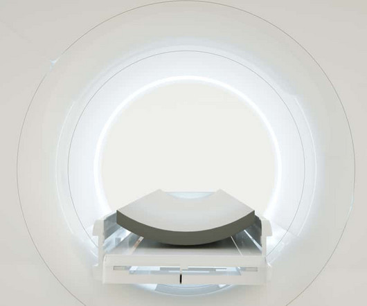
Diagnostic imaging includes different types of scans that take pictures of the inside of the body. Some of the most common types are: X-rays : Used to check for broken bones, lung infections, and more. MRI: Uses magnets and radio waves to create detailed images of tissues and organs. What is Diagnostic Imaging?
tesla MRI AI body composition analysis Cardiac PET Cryo/thermoablation CT colonography Genicular artery embolization Hyperpolarized xenon-129 MRIPET/MRI Photon-counting CT Radiomics Theranostics Whole-body MRI screening Image of the Year 3D PET/MR image. tesla brain MRIscans.
Medical imaging has evolved over centuries, starting with X-rays in 1895, progressing to CT, MRI, and PETscans. The post Revolutionising Healthcare: A Historical Perspective on Medical Imaging appeared first on Open Medscience.
Reimbursement barriers Unfortunately, until recently, there were significant barriers to receiving amyloid PET imaging in some regions. Centers for Medicare and Medicaid Services (CMS), for example, did not cover amyloid PETscans outside of clinical trials for Medicare beneficiaries and only covered one scan per patient’s lifetime.
The answer to many of these questions could be just one—MRI. Offer Versatility and Clarity to Your Doctors and Patients MRI, or Magnetic Resonance Imaging , could be the most versatile imaging technology available to members of the medical community. However, this is also the case with virtually any other form of diagnostic imaging.
To help guide the diagnostic process, the following imaging procedures may be used: X-Ray: Typically the first requested imaging procedure, X-rays help identify respiratory abnormalities and other lung issues. This technology can indicate a thickened pleura, fluid between the lungs and chest wall and calcium deposits.
The Impact of Imaging in Diagnosis: Explore traditional imaging methods such as X-rays, CT scans, and MRIs in diagnosing sarcoma and bone cancer. Understanding Sarcoma and Bone Cancer: Provide a brief overview of sarcoma and bone cancer, emphasizing the rarity and complexity of these types of cancers.
This procedure is less invasive than other methods and provides a higher level of detail compared to traditional X-rays, while also reducing radiation exposure. CT Scan-Guided Biopsy: This technology better identifies the location of the growth, so that a sufficient number of cells can be gathered from the tumor for analysis.
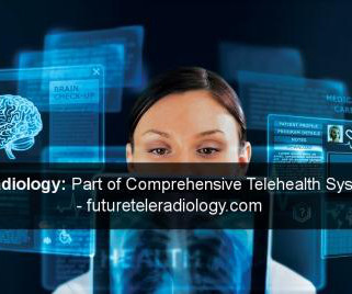
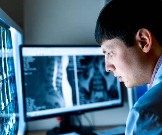
Diagnosis often depends on the interpretation of tests like of X-Rays, CT Scan, MRI, PET CT Scan , etc. It has adhered to the globally standardized reporting format of X-Rays, CT Scan, MRI, PET CT Scan, etc. Such situations were created in telehealth.
PACS works as a host that integrates the radiological images acquired from different radiological imaging modalities (X-Ray, Ultrasound, CT, MRI , PETScan, Nuclear Medicine, etc…) with a network of information system (RIS and or HIS), EMR, different work stations and image storage/archival system.
But, first, let’s understand what a PET/CT is and why it is used. What is a PET/CT? A PET/CT is the combination of PET (Positron Emission Tomography) and CT ( Computed Tomography ) scans. A PETscan creates images of your body’s biological processes. The PET shows where the tracer is in your body.
This standard has revolutionized the radiology industry, encompassing many imaging modalities such as X-rays, computed tomography (CT), magnetic resonance imaging (MRI), ultrasound, nuclear medicine, PETscans, etc.
We organize all of the trending information in your field so you don't have to. Join 5,000 users and stay up to date on the latest articles your peers are reading.
You know about us, now we want to get to know you!
Let's personalize your content
Let's get even more personalized
We recognize your account from another site in our network, please click 'Send Email' below to continue with verifying your account and setting a password.


Let's personalize your content