This site uses cookies to improve your experience. To help us insure we adhere to various privacy regulations, please select your country/region of residence. If you do not select a country, we will assume you are from the United States. Select your Cookie Settings or view our Privacy Policy and Terms of Use.
Cookie Settings
Cookies and similar technologies are used on this website for proper function of the website, for tracking performance analytics and for marketing purposes. We and some of our third-party providers may use cookie data for various purposes. Please review the cookie settings below and choose your preference.
Used for the proper function of the website
Used for monitoring website traffic and interactions
Cookie Settings
Cookies and similar technologies are used on this website for proper function of the website, for tracking performance analytics and for marketing purposes. We and some of our third-party providers may use cookie data for various purposes. Please review the cookie settings below and choose your preference.
Strictly Necessary: Used for the proper function of the website
Performance/Analytics: Used for monitoring website traffic and interactions
Some of my radiological heroes would report a staggering 30,000 to 40,000 radiographs a year. So as to report more CT and MRI, radiologists stopped doing hands-on ultrasound and fluoroscopy. Some even [startled gasp] gave up reporting plain radiographs. Many diagnostic radiologists became pure CT and MRI specialists.
MRI safety articles have always been popular with our members, so it’s not surprising that our top article in 2019 reported on a safety incident in Sweden. The police investigation into last Wednesday's accident at a mobile MRI unit in Swedish Lapland is underway and likely to take several weeks.
A team led by Yin Ting Chiu, PhD, from the Hong Kong Children’s Hospital discussed key factors identified by the hospital’s MR team for effectively performing supplementary MRI scans on children ages three to seven without sedation. Performing MRI scans on young children can be challenging for radiologists.
Are low-field MRI units effective for neuroradiologic imaging? Use of low-field MRI for this indication would improve patient care by expanding access to the modality, wrote a team led by medical student Lauren Kelsey. The group's commentary was published February 13 in RadioGraphics. tesla is lower than that at 1.5-tesla,
MRI helps clinicians assess the neural involvement in endometriosis and could help them prevent irreversible nerve damage and chronic sensorimotor neuropathy in women suffering from the condition, Cleveland Clinic researchers have reported. The team's review of MRI's role for this indication was published January 3 in RadioGraphics.
The first scans have been performed in the Olympic imaging polyclinic ahead of Friday's opening ceremony, and the 68-strong squad of radiologists and radiographers are primed and ready for action, according to musculoskeletal (MSK) expert Jérôme Renoux, MD. One of the two MRI scanners located in trucks in the Olympic Village.
Its impact on radiographer workflow ranges from detecting poor image quality on x-ray; automating CT imaging protocols; and for MRI, streamlining workflows for faster scan times, image reconstruction, and using synthetic MRI sequences.
Middlebrooks' research interest consists of using ultrahigh-field, 7-tesla MRI to plot brain microstructure and develop surgical treatment of brain tumors, epilepsy, and neurodegenerative and movement disorders such as Parkinson's disease, essential tremor, and dystonia. I'm a radiographer,' " Stewart recalled.
AI highlights clinically significant prostate cancer on MRI Sunday, November 26 | 9:30 a.m.-9:40 S1-SSGU01-3 | Room S404 This session features an update on research toward clinical translation of AI in detecting clinically significant prostate cancer on MRI. 9:40 a.m. | 10:00 a.m. | 1:50 p.m. | 3:50 p.m. | 10:20 a.m. | 3:10 p.m. |
SINGAPORE - Does MRI have a role in the emergency department (ED)? MRI complements CT, ultrasound, and x-ray when it comes to trauma imaging," Mandel said. And having MRI available in the emergency department means that ED patients don't have to compete with inpatients or outpatients for MRI time."
At the time, there was little training for, say, cardiac MRI. Together with our radiographers, I learned to scan cardiac patients and learned special anatomy from pediatric cardiologists and pediatric cardiac surgeons." Gutberlet described how he came to cardiac imaging early in his medical career. But he persevered.
Current MRI definitions of knee osteoarthritis aren't necessarily adequate for identifying which knees will develop disease in the future, researchers have found. However, a substantial proportion of individuals with baseline MRI-defined knee OA did not develop radiographic knee OA during follow-up." times increased risk.
The green project conducted by radiographers at the European Institute of Oncology in Milan and nearby Hospital of Legnano saved an estimated 12,000 euros ($13,000), said Andrea Masperi, who presented the details. during indirect radiology department activities, according to findings of a pilot study presented March 1 at ECR 2024.
When reviewing radiographs, computed tomography (CT) scans or magnetic resonance imaging (MRI) scans, do you still turn to mnemonics every now and then to jog your short-term memory?
His research interests include using structural and functional MRI -- particularly ultrahigh-field, 7-tesla MRI -- to map brain microstructure and develop neurosurgical treatment of brain tumors, epilepsy, and neurodegenerative and movement disorders such as Parkinson's disease, essential tremor, and dystonia.
A newly developed Musculoskeletal Infection-Reporting and Data Systems (MSKI-RADS) framework is effective when it comes to standardized terminology and recommended management of MRI findings of extremity infections, researchers have reported. But a consistent interpretation system for MRI results has been lacking.
Xray- Frontal radiograph of wrist shows multiple osteolytic areas with surrounding sclerosis is seen involving all carpal bones and second metatcarpal bone s/o permeative type of bone destruction.
Lack of AI knowledge among educators was the top reason for not integrating AI in education," noted a team led by MRIradiographer Nikolaos Stogiannos of the University of London in the U.K. In a survey of all U.S. The study was published July 13 in the Journal of Medical Imaging and Radiation Sciences.
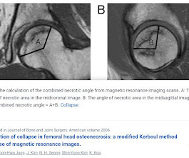
MRI study of hip joints shows: Avascular necrosis involving bilateral capital femoral epiphysis. Modified Kerboul method is used for prediction of collapse in femoral head osteonecrosis by volumetric analysis on MRI. HOW IS IT CALCULATED ? With use of the modified method of Kerboul et al.,
milla1cf Fri, 02/16/2024 - 10:13 February 16, 2024 — Fujifilm Healthcare Europe ipresented the Echelon Synergy – a revolutionary MRI scanner for efficient, high performance imaging with optimal patient comfort – at ECR 2024. The Echelon Synergy MRI scanner also provides benefits to boost the efficiency of workflows.
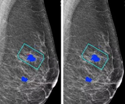
For the AI study, two views of each breast (four images) are taken to be evaluated independently either by the AI system plus a single reader or by a radiologist/radiographer as a first reader and second reader using the two-reader model. when using an AI system to differentiate cancers from benign lesions at breast MRI.
. | S1-SSCH01-5 | E451A This scientific paper may increase overall confidence in the potential of using multimodal AI for tuberculosis (TB) detection, and potentially autonomous reporting, on chest radiographs in certain clinical settings. 3D body composition analysis of whole-body MRI can predict mortality Wednesday, December 4 | 10:06 a.m.-10:18
A team of 32 radiologists and 36 radiographers are limbering up to work at the summer Olympics, which begins July 25. The imaging department at the polyclinic will be equipped with two mobile MRI scanners (Ingenia, from Philips), three ultrasound machines (Aplio i800, from Canon), and one x-ray system (from Primax International).
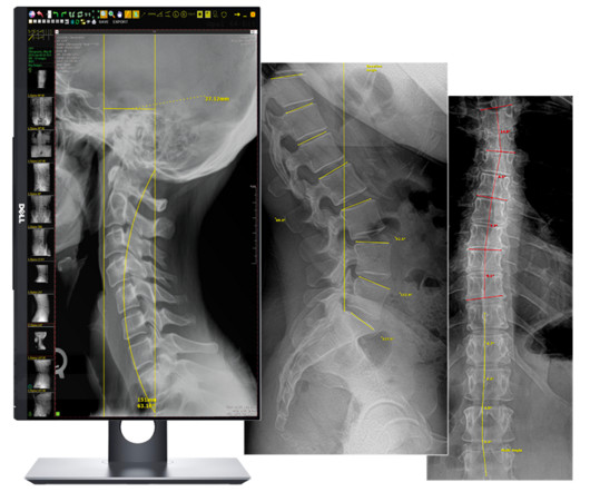
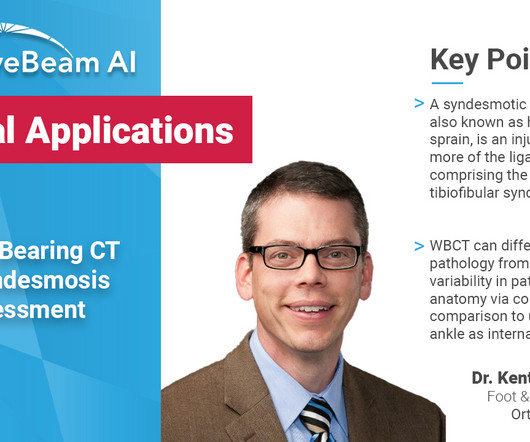
Key Points: Currently plain radiographs are the standard method in diagnosing syndesmotic ankle injuries even though the distal tibiofibular joint cannot be assessed due to superposition of the osseous structures in the foot.
The first female editor in chief of RSNA’s peer-reviewed RadioGraphics journal since 2021, Menias splits her time between Mayo Clinic, where she’s a specialist in abdominal imaging, and the journal, where she is achieving her first goals of expanding RadioGraphics as a learning platform. And they're going to chip away at waste."
For CT and MRI procedures where injected contrast media is ordered, ". Virtual supervision of RTs In Alabama, providers have had questions about RT supervision, leading the Alabama State Board of Medical Examiners to consider declaring a position on what constitutes direct supervision by a board-certified radiologist.
Qureshi said these findings highlight the need to reduce the reliance on radiographic media contrast imaging without compromising patient outcomes. Some suggested methods include doing more magnetic resonance imaging ( MRI ) scans and reducing the amount of dye used per patient.
Moreover, survey responses after the course indicated average student confidence increased by more than one point on a six-point Likert scale in radiographic interpretation (p = 0.004), ultrasound interpretation (p = 0.0002), CT/MRI interpretation (p = 0.02), general radiology knowledge including procedural skills (p = 0.0001), and appropriate image (..)
MRIs and CT scans are advanced medical imaging techniques that create detailed images of the internal structures of the body. These enable a wide range of diagnostics for conditions that can often not be diagnosed using traditional radiographic imaging techniques.
The approval expands upon Bayer's focus on breast imaging, with a portfolio that also includes Gadavist (gadobutrol) injection, a gadolinium-based contrast agent approved for use with MRI ( Magnetic Resonance Imaging ) to assess the presence and extent of malignant breast disease in adult patients. RadioGraphics 2019 39:7, 1907-1920.
Patients with ARIA sometimes have headaches, but they are usually asymptomatic and only diagnosable with MRI. “It Most patients with asymptomatic ARIA meeting specific radiographic and clinical criteria may continue to receive treatment. It is essential for the radiologist to recognize and monitor ARIA,” Dr. Agarwal said. “As
The enduring shortage is affecting staffing for radiographers and radiologists; and all imaging modalities. Executive leadership also plays an essential role in retention of radiology staff, emphasized Keith Aldahondo, MHA, RT (CT) (MR) CRA, CT/MRI, Manager at Tampa General Hospital in Florida. “We Bureau of Labor Statistics.
For dense-breasted patients requiring supplemental imaging, MRI remains a valuable option that is not limited by breast density and is shown to be more sensitive than mammography at finding breast cancer. The shortage of radiographers: A global crisis in healthcare. J Med Imaging Radiat Sci. 2023 Oct 19:S1939-8654(23)01877-5.
Kim Mason Kim Mason, an Audit and Research Radiographer for Mid Yorkshire Teaching Hospitals Trust, talks about their role as well as the value of radiographer engagement in research activities and how to get involved. Hi, I’m Kim and I am an alternative-styled, funky-haired, septum-pierced, disabled Audit and Research Radiographer.
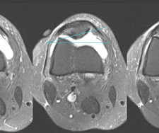
This MRI Axial STIR sections of knee show clinical marker on skin on anteromedial aspect of knee joint. However, lateral tilt of patella on axial sections of MRI or sunrise knee radiographs, patella facing medially without lateral translation should be depicted meticulously which is very commonly overlooked during MRI interpretation.
Positions (On-site): Body (100% Body) – Regions Hospital Mix of shifts worked on-site Mixture of hospital, outpatient, and remote Interpret MRI, CT, U/S, and radiographs After-hours coverage provided internally by the emergency radiology section No neuro or MSK Body/Mammo – Western Wisconsin 45-minute drive from the Twin Cities.
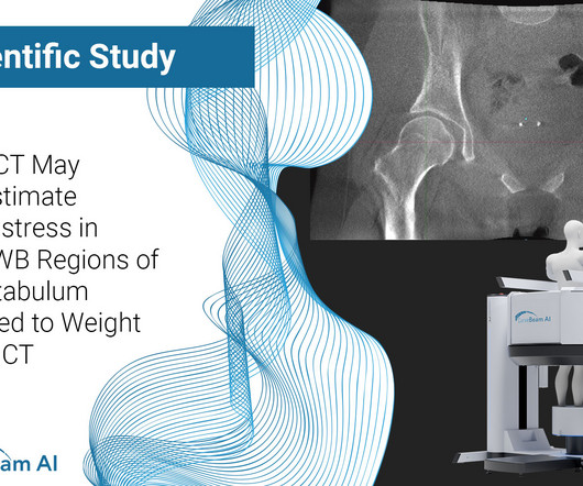
Knee osteoarthritis (OA) clinical trials results show that weight bearing CT (WBCT) imaging may offer new insights into OA pathology beyond what plain radiographs and MRI can provide. Demonstrates higher sensitivity and accuracy for detecting osteophytes and subchondral cysts in comparison to fixed flexion radiographs.
Computational models of living patients are typically generated from clinical NWB supine CT or MRI scans, which require scans to be reoriented to a neutral, standardized pose before functional load application. The reorientation process involves translating the 3D models to a standardized coordinate system of bone landmarks.
Cardiac CT and MRI Needs Are Increasing – Are You Ready? Changes in the standard of care for cardiac imaging means there’s an increasing need for radiologists to be able to read cardiac CT and MRI. AMA PRA Category 1 Credits Focus Your Training with our Cardiac MRI and CT Library Plan!
Education/Requirements: Graduate of an accredited program in Radiological Technology ARRT registered, MR certified Florida state license for General Radiographer Valid driver's license Work Schedule: The schedule for this position will be every weekend, Saturdays and Sundays, 12-hour shifts guaranteed 2 paid weekends of the employee's choice, non-consecutive (..)
Dr. Lee’s clinical and scientific interests include abdominal and pelvic imaging, women’s imaging , radiation safety, advanced MRI and CT , molecular imaging and gynecological cancers. She has served on the RSNA Genitourinary Scientific Program Committee and RadioGraphics Genitourinary Review Panel.
A weight bearing CT scan can: Provide increased sensitivity and specificity over radiographs. WBCT scans can also be used as an adjunct to an ankle MRI to correlate anatomic abnormalities with functional instability. Diagnosis If WB X-rays are indeterminate after clinical exam, order a bilateral WBCT.
MRI is key to diagnosis, obtain this imaging in all patients who raise clinical suspicion Patients with hemodynamic instability and neurologic compromise warrant empiric antibiotics. Imaging Gadolinium enhanced MRI – modality of choice, highly sensitive and specific ( Mylona 2009 ). Read more
Key Points: Weight bearing CT (WBCT) can detect signs of osteoarthritis (OA), such as osteophytes, subchondral cysts, and joint space narrowing better than radiographs. Emerging evidence suggests WBCT reveals meniscal extrusion not detected by MRI and WBCT arthrography visualizes meniscal tears. K.Kaneda et al. Segal et al.
We organize all of the trending information in your field so you don't have to. Join 5,000 users and stay up to date on the latest articles your peers are reading.
You know about us, now we want to get to know you!
Let's personalize your content
Let's get even more personalized
We recognize your account from another site in our network, please click 'Send Email' below to continue with verifying your account and setting a password.


Let's personalize your content