This site uses cookies to improve your experience. To help us insure we adhere to various privacy regulations, please select your country/region of residence. If you do not select a country, we will assume you are from the United States. Select your Cookie Settings or view our Privacy Policy and Terms of Use.
Cookie Settings
Cookies and similar technologies are used on this website for proper function of the website, for tracking performance analytics and for marketing purposes. We and some of our third-party providers may use cookie data for various purposes. Please review the cookie settings below and choose your preference.
Used for the proper function of the website
Used for monitoring website traffic and interactions
Cookie Settings
Cookies and similar technologies are used on this website for proper function of the website, for tracking performance analytics and for marketing purposes. We and some of our third-party providers may use cookie data for various purposes. Please review the cookie settings below and choose your preference.
Strictly Necessary: Used for the proper function of the website
Performance/Analytics: Used for monitoring website traffic and interactions
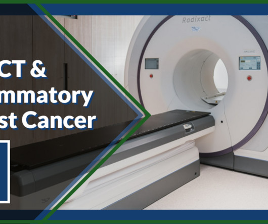
Computerized tomography (or a CT scan) combines images from different angles to create cross-section pictures of the body with more information than typical X-ray technology. Positron Emission Tomography (a PETscan) uses nuclearmedicine to detect metabolic signals emitted by cancer cells.
Flavio Forrer , MD, PhD, Chairman of NuclearMedicine in the Division of Radiology and NuclearMedicine at Kantonsspital St. GE HealthCare’s deep-learning-enabled software is revolutionizing image acquisition and reconstruction in MR, CT , X-ray and now PET/CT, empowering clinicians and helping improve patient outcomes.
To help guide the diagnostic process, the following imaging procedures may be used: X-Ray: Typically the first requested imaging procedure, X-rays help identify respiratory abnormalities and other lung issues. This technology can indicate a thickened pleura, fluid between the lungs and chest wall and calcium deposits.
PACS works as a host that integrates the radiological images acquired from different radiological imaging modalities (X-Ray, Ultrasound, CT, MRI , PETScan, NuclearMedicine, etc…) with a network of information system (RIS and or HIS), EMR, different work stations and image storage/archival system.
This standard has revolutionized the radiology industry, encompassing many imaging modalities such as X-rays, computed tomography (CT), magnetic resonance imaging (MRI), ultrasound, nuclearmedicine, PETscans, etc.
tesla MRI AI body composition analysis Cardiac PET Cryo/thermoablation CT colonography Genicular artery embolization Hyperpolarized xenon-129 MRI PET/MRI Photon-counting CT Radiomics Theranostics Whole-body MRI screening Image of the Year 3D PET/MR image. PETscans predict patient response to Pluvicto.
Reimbursement barriers Unfortunately, until recently, there were significant barriers to receiving amyloid PET imaging in some regions. Centers for Medicare and Medicaid Services (CMS), for example, did not cover amyloid PETscans outside of clinical trials for Medicare beneficiaries and only covered one scan per patient’s lifetime.
We organize all of the trending information in your field so you don't have to. Join 5,000 users and stay up to date on the latest articles your peers are reading.
You know about us, now we want to get to know you!
Let's personalize your content
Let's get even more personalized
We recognize your account from another site in our network, please click 'Send Email' below to continue with verifying your account and setting a password.


Let's personalize your content