This site uses cookies to improve your experience. To help us insure we adhere to various privacy regulations, please select your country/region of residence. If you do not select a country, we will assume you are from the United States. Select your Cookie Settings or view our Privacy Policy and Terms of Use.
Cookie Settings
Cookies and similar technologies are used on this website for proper function of the website, for tracking performance analytics and for marketing purposes. We and some of our third-party providers may use cookie data for various purposes. Please review the cookie settings below and choose your preference.
Used for the proper function of the website
Used for monitoring website traffic and interactions
Cookie Settings
Cookies and similar technologies are used on this website for proper function of the website, for tracking performance analytics and for marketing purposes. We and some of our third-party providers may use cookie data for various purposes. Please review the cookie settings below and choose your preference.
Strictly Necessary: Used for the proper function of the website
Performance/Analytics: Used for monitoring website traffic and interactions
Some of my radiological heroes would report a staggering 30,000 to 40,000 radiographs a year. Some even [startled gasp] gave up reporting plain radiographs. And they did this alarming juggling act for hour after hour, day after day, demolishing huge piles of film packets. I still don’t know how they did it.
Shamie Kumar describes how AI fits into a radiology clinical workflow and her perspective on how a clinical radiographer could use this to learn from and enhance their skills. If the AI findings are seen in PACS, how many radiographers actually log into PACS after taking a scan or X-ray? Can Radiographers Up-Skill?
milla1cf Wed, 06/12/2024 - 21:59 June 12, 2024 — Carestream launched its Image Suite MR 10 Software to help deliver a boost to productivity and efficiency while enabling a more user-friendly imaging experience for radiographers. Image Suite MR 10 helps push the envelope further for even more focus on patient care.”
Commercially Available Chest Radiograph AI Tools for Detecting Airspace Disease, Pneumothorax, and Pleural Effusion. Generative Artificial Intelligence for Chest Radiograph Interpretation in the Emergency Department. Cognitive Motor Dissociation in Disorders of Consciousness. To learn more about this paper, click here.
Carestream’s affordable DR Retrofit solution is comprised of our Focus 35C/43C Detectors, ImageSuite software, an optional mini-PACS, and DRYVIEW Laser Imagers for a complete, fully digital capture-to-print solution. We also offer an optional and affordable mini-PACS module that increases productivity and patient care even more.
Introduction: In the digital era of healthcare, Picture Archiving and Communication Systems (PACS) have emerged as orchestras orchestrating the seamless management and communication of medical images. Efficient Image Storage and Retrieval: Explore how PACS facilitates efficient storage and retrieval of medical images.
Imagine that your radiology department has no access to your PACS, your RIS, or even email. Our radiology department was one of the fortunate ones, we were without PACS “only” six days. Other Irish healthcare sites were without PACS for almost five weeks. No PACS/RIS. How would you create and distribute reports?
In the third blog of her series on AI and the radiographer, Shamie Kumar explores the impact on the radiographer when AI is integrated within an imaging modality. The question to explore in this blog is when AI is integrated within an imaging modality itself and how that may impact a radiographer.
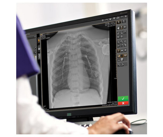
A fundamental goal of radiographers is to complete an imaging exam that provides sufficient information for an accurate clinical diagnosis–and at the lowest possible dose. We also have a Detector Verification alert that signals radiographers when they choose the wrong detector–which would result in the need to repeat the exam.
Physicians have better diagnostic confidence when they can view radiographs in the manner most suitable to their preferences. Your radiographers need to work as efficiently as possible in order to keep pace. This makes it easier for radiographers to interact with different DR systems; and it reduces training time and costs.
Written ostensibly for radiographers/ radiology technologists, this 200-page soft cover book, written by Quinn Carroll, RT and now in its second edition, contains basic and important information not only for technologists but also for radiologists——- residents and practicing radiologists. Carroll QB.
The implementation of Picture Archiving and Communication System (PACS), and rising R&D efforts have increased the need for teleradiology facilities. Orthopantamogram (OPG), DentaScan (Dental CT), Intra-Oral Radiographs (Periapical/Occlusal/Bitewing), Cephalogram Tracing and Analysis, Sialogram, etc.
Note that everyone’s combination of PACS, dictation software, and personal preferences is a little different, so it really is worth spending ten minutes to make sure you have the combination of features you want (and ideally the understanding of how to adjust them for troubleshooting).
Any time of the day or night, a clinician, radiographer, or radiology manager can call TMC to discuss scanning a patient. For elective services, the AI software looks for PEs in all CT examinations that involve the thorax as soon as the examination arrives in the TMC PACS.
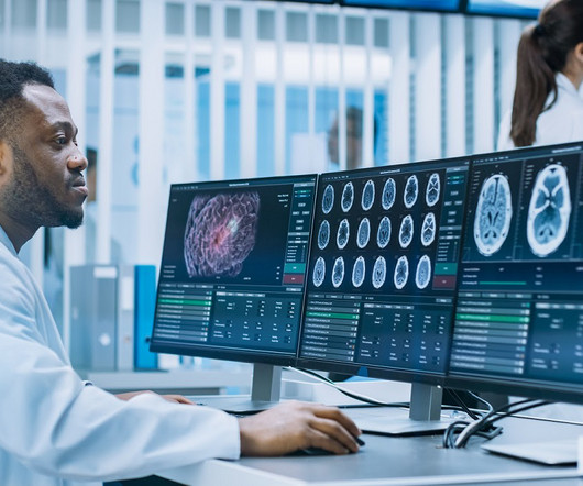
Here are some ways the technology is transforming workflows in fields like radiology, cardiovascular care, neurology and emergency medicine: Radiology In radiology, AI excels at recognizing complex patterns in imaging data and providing quantitative assessments of radiographic characteristics.
Various software (PACS & DICOM standards) and hardware support the whole system in maintaining the quality of the image during the process (acquisition, transmission, storage, retrieval); and different compression techniques are employed to contain huge data and maintain the integrity of the digital images.
Photoprint from radiograph by W.K. 3) In the early twentieth century, it was a common goal for investigators to try to find a way to separate the superimposed shadows that were recorded when a complex structure was shown on a radiograph. (3) This is now known as ‘Hand mit Ringen’. (1) after seeing the image. (2) Röntgen, 1895.
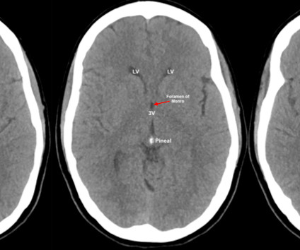
This can be done manually or, typically, PACS viewers have preset brain, bone, lung and soft tissue windows that can be displayed by pressing different numbers on the keypad. RadioGraphics, 1998; 18(1):151-163 3. Computed Tomography. In Grainger & Allison's Diagnostic Radiology, 6th Edition (2016). McKetty MH. X-ray attenuation.
A PACS-integrated AI tool not only correctly identified pneumothorax on inpatient chest x-rays but also prioritized scans and improved radiologist reporting times, according to a group in Cleveland, OH.
We organize all of the trending information in your field so you don't have to. Join 5,000 users and stay up to date on the latest articles your peers are reading.
You know about us, now we want to get to know you!
Let's personalize your content
Let's get even more personalized
We recognize your account from another site in our network, please click 'Send Email' below to continue with verifying your account and setting a password.


Let's personalize your content