This site uses cookies to improve your experience. To help us insure we adhere to various privacy regulations, please select your country/region of residence. If you do not select a country, we will assume you are from the United States. Select your Cookie Settings or view our Privacy Policy and Terms of Use.
Cookie Settings
Cookies and similar technologies are used on this website for proper function of the website, for tracking performance analytics and for marketing purposes. We and some of our third-party providers may use cookie data for various purposes. Please review the cookie settings below and choose your preference.
Used for the proper function of the website
Used for monitoring website traffic and interactions
Cookie Settings
Cookies and similar technologies are used on this website for proper function of the website, for tracking performance analytics and for marketing purposes. We and some of our third-party providers may use cookie data for various purposes. Please review the cookie settings below and choose your preference.
Strictly Necessary: Used for the proper function of the website
Performance/Analytics: Used for monitoring website traffic and interactions
While radiographers are concerned about job security, they are also optimistic about AI’s role in their future workflows, according to a presentation given March 1 at ECR 2024. Radiographers appear optimistic about the future of radiographer job roles and responsibilities,” Walsh said. Of the total respondents, 31.3%
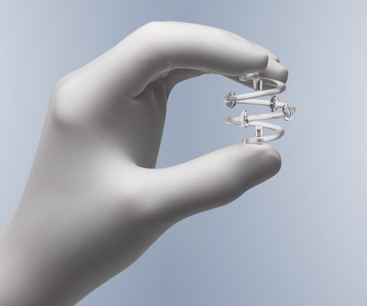
The alert pertains to the BioZorb and BioZorb LP radiographic markers, implanted in soft tissue to indicate the site for radiation therapy or other medical procedures.
ChatGPT can answer patient questions about radiation protection for medical imaging exams comparably to websites of radiology institutions, according to research published June 25 in Radiology. Median scores of answers to patient questions on radiation protection in radiology Radiology institutional websites ChatGPT Scientific adequacy 5.4
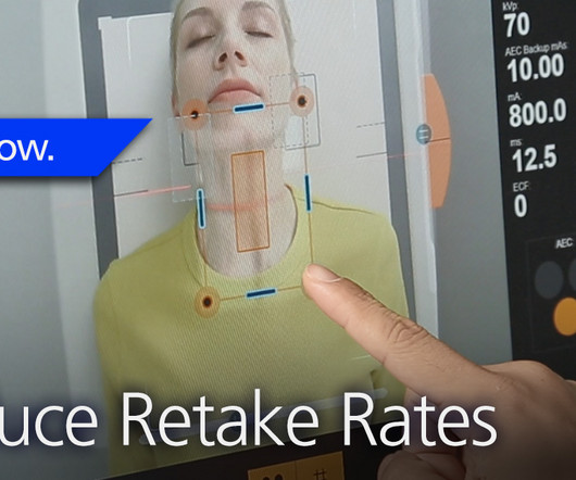
At RSNA 2023, look for AI-driven systems that radiographers can use to help make patient positioning faster and more precise, and bring consistency to the process, all of which help improve image quality and reduce the need for retakes. Even the most skilled radiographers can fail to get positioning just right.
Staff operating ionizing radiation equipment in Michigan are now required to meet certain qualifications for active status and employment. The rules also establish initial and continuing education requirements for limited-scope radiographers and radiologist assistants. Read the full filing here.
Food and Drug Administration (FDA) for not addressing violations related to the companys BioZorb 3D bioabsorbable radiographic markers. Hologic has received a warning letter from the U.S.
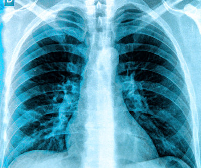
Spirometry values were below predicted values and a standard chest radiograph depicted an elevated right hemidiaphragm that was not present on a prior CT examination during his SARS-CoV-2 infection. With the dynamic functional imaging capability of DDR, the authors could visualize thoracic and pulmonary motion and track diaphragm movement.
a) Raw example of a dynamic digital radiograph. (b) The digital technology limits radiation exposure to patients compared with standard chest x-rays, they wrote. To that end, the group developed two convolutional neural networks (CNN) designed to quantify key measurements in DDR image sequences.
DDR is an emerging imaging technique that uses a pulsed x-ray source to acquire a series of radiographs at six to 15 frames per second. These images are then processed to visualize joints in motion.
Shamie Kumar describes how AI fits into a radiology clinical workflow and her perspective on how a clinical radiographer could use this to learn from and enhance their skills. If the AI findings are seen in PACS, how many radiographers actually log into PACS after taking a scan or X-ray? Can Radiographers Up-Skill?
Communication among practitioners: Effective communication between radiographers, radiologists, and child-life specialists is needed to discuss scan protocols, streamline the exam, and minimize table time. Having support from a child-life specialist inside the scan room can help children stay calm and cooperative.
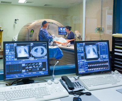
“So we were thinking and asking ourselves, ‘can nonradiologists benefit from AI and chest radiography analysis in this emergency unit set.’ ” Per year, LMU receives between 5,000 and 6,000 orders for chest radiographs for primary diagnosis from the emergency unit alone.
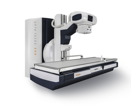
Additionally, all table movements can be controlled by the radiographer from the touch screen Automatic positioning of all table axes depending on the selected exam Flexibility to control table movements with foot pedals, a remote, or from the console, depending on the user’s preference In addition, DRX-Excel Plus includes a camera integrated into (..)
"Lack of AI knowledge among educators was the top reason for not integrating AI in education," noted a team led by MRI radiographer Nikolaos Stogiannos of the University of London in the U.K. The study was published July 13 in the Journal of Medical Imaging and Radiation Sciences.
Commercially Available Chest Radiograph AI Tools for Detecting Airspace Disease, Pneumothorax, and Pleural Effusion. Effects of low-dose ionizing radiation on genomic instability in interventional radiology workers. Generative Artificial Intelligence for Chest Radiograph Interpretation in the Emergency Department.
Whenever bilateral standing radiographs would have been needed, a WBCT was performed instead. The reality is that if insurance was not an issue, I would not perform conventional radiographic imaging anymore. WBCT imaging allows superior and more complete 3-dimensional assessment of the foot and ankle, with low radiation dose.
She currently serves as chair of the department of medical imaging and radiation sciences in the College of Health Professions at TJU and directs the radiography and invasive cardiovascular technology programs. Her department currently serves 109 students who are studying in one of the university's nine concentration options.
Detecting CSIs in a clinical setting often requires imaging such as X-rays and computed tomography (CT) scans, both of which expose children to radiation, which can cause other health issues over time.
Teleradiology-India Introduction: “X-ray Visionaries” takes you on a compelling journey to unveil the expertise of radiographers and technologists, the unsung heroes of X-ray technology. Chapter 1: Introduction to Radiographers and Technologists An overview of the pivotal roles radiographers and technologists play in healthcare.
The American Society of Radiologic Technologists (ASRT) has partnered with medical imaging professional and actor, Michael Benzaia, for its new "Be Seen" campaign to raise public awareness about the role of medical imaging and radiation therapy professionals in patient diagnosis, intervention, and treatment.
In an open forum for radiographers at ECR 2024, Yi Xiang Tay, of Singapore University Hospital's radiography and diagnostic imaging department, shared his team's systematic review of the impact of imaging referral guidelines on patients and radiology services.
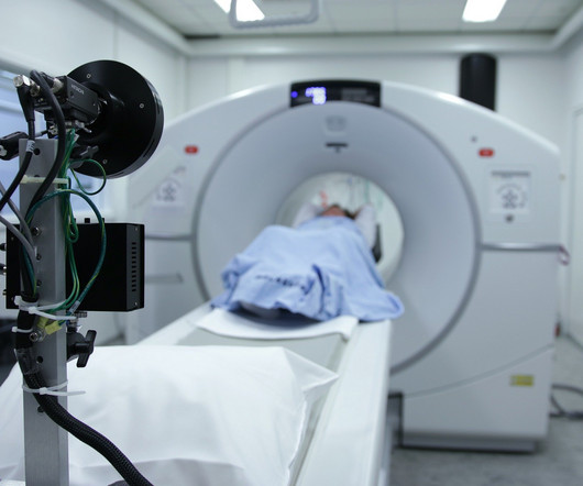
In a new study, a radiograph device equipped with an AI-powered deep learning neural network has demonstrated improved image quality in pediatric X-rays.
Repeating imaging exams increases the workload of your radiographers who are already stretched too thin; increases the exposure of the affected patients; and contributes to patients’ reduced confidence and satisfaction with your imaging department. The Audio Assist makes it easier for radiographers to hear the patients.
Additionally, all table movements can be controlled by the radiographer from the touch screen, enabling easy setup of either fluoroscopy or radiography settings. In addition to improving workflow, the DRX-Excel Plus X-ray System has new features to help limit the radiation dose.
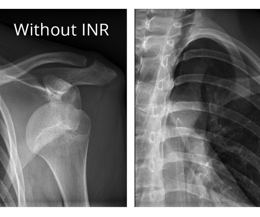
The company will showcase the clinical analysis of Canon’s Intelligent Noise Reduction (Intelligent NR) that provides superior image quality while lowering radiation dosing in pediatric digital radiography at the Radiological Society of North America Annual Meeting 2023 (RSNA) , McCormick Place Convention Center, Chicago, IL Nov.
Geoffrey Rubin, MD, of the University of Arizona College of Medicine Tucson, offered session attendees an overview of key issues in the radiology workforce landscape, highlighting a 2024 consensus committee report from the American Society of Radiologic Technologists (ASRT) on the future of medical imaging and radiation therapy.
The World Health Organization (WHO) on September 17 will offer a webinar on the importance of accurate and timely diagnoses in ensuring patient safety that will include discussion of medical imaging and radiation exposure. to 1:30 p.m. to 1:30 p.m.
The ASRT weighed in, telling AuntMinnie.com, "A key issue we continue to face is state legislation designed to weaken or remove licensure for medical imaging and radiation therapy professionals.
A fundamental goal of radiographers is to complete an imaging exam that provides sufficient information for an accurate clinical diagnosis–and at the lowest possible dose. We also have a Detector Verification alert that signals radiographers when they choose the wrong detector–which would result in the need to repeat the exam.
But at RSNA 2023, part of the focus was on AI advances that are changing how radiographers work today—for example, continuing to help us balance the goal of capturing the most information possible in an image without excessive radiation dose. AI tools that enable radiographers to separate noise from an image already exist today.
A study published in the Journal of Medical Radiation Sciences compared 270 dental X-rays, from people who were told to hold their tongues against the roof of their mouths throughout the procedure.
milla1cf Fri, 02/23/2024 - 10:22 February 23, 2024 — The American Society of Radiologic Technologists (ASRT) launched its "Be Seen" campaign today to raise public awareness about the crucial role medical imaging and radiation therapy professionals play in patient diagnosis, intervention and treatment.
Alternatively, DCR, also referred to as dynamic digital radiography, is a low ionizing radiation x-ray system that produces wide field-of-view fluoroscopic-style images of the thorax in motion. Yet FEV1 may not always reflect the severity of the airway obstruction, they noted.
Other modalities like CT are not portable, bring increased radiation exposure, in addition to risks associated with intra-hospital patient transportation. Our radiographic spectral images separate materials such as water (i.e., soft tissue, lung lesions etc.) and calcium (i.e.
Kim Mason Kim Mason, an Audit and Research Radiographer for Mid Yorkshire Teaching Hospitals Trust, talks about their role as well as the value of radiographer engagement in research activities and how to get involved. Hi, I’m Kim and I am an alternative-styled, funky-haired, septum-pierced, disabled Audit and Research Radiographer.
CBCT is an innovative imaging modality with a body of supporting literature reporting reduced radiation exposure, shortened examination time and operating time, and a decreased time interval between injury and diagnosis. Researchers analyzed the limited literature available on using CBCT as a primary modality for diagnosing AS and CLAI.
Alessio Bernasconi, MD, PhD, FEBOT Azienda Ospedaliera Universitaria Federico II of Naples Department of Orthopaedic Surgery Naples, Italy I think that every condition for which one would traditionally request a bilateral standing radiographic imaging of both feet is a good indication for WBCT. Even soft tissue issues (i.e.
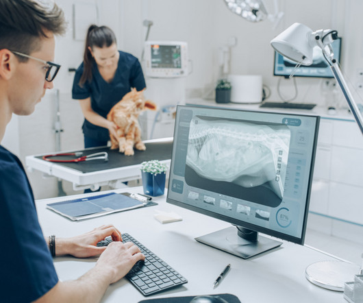
Veterinary technicians play a pivotal role in the radiographic process within veterinary practices. Poor positioning can lead to retakes, increased radiation exposure, and misdiagnoses. Safety Protocols Radiation Safety Protecting both the veterinary staff and the animals from unnecessary radiation exposure is paramount.
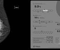
Transforming the breast dose model The prestigious Joint AAPM Task Group 282/EFOMP Working Group, focused on modernizing breast radiation dose modeling, recently published a report outlining their new model in Medical Physics. Volumetric breast density emerges as an essential input to facilitate patient-based radiation dose estimates.
X-ray Also called a radiograph, an X-ray uses radiation to create images of the body. Like the X-ray, a CT scan sends radiation through your body with a much detailed view of its structures. Unlike X-rays, MRIs don’t have the harmful effects of radiation.
How are radiographers treated when they admit to making a mistake? Incorrect markers can result in a patient having to undergo a second exam and additional exposure to radiation. Some safety incidents have nothing to do with radiation. Two, periodically review with radiographers the process for reporting safety incidents.
Dr. Lee’s clinical and scientific interests include abdominal and pelvic imaging, women’s imaging , radiation safety, advanced MRI and CT , molecular imaging and gynecological cancers. She has served on the RSNA Genitourinary Scientific Program Committee and RadioGraphics Genitourinary Review Panel.
Lumbosacral spine X-rays, also called lumbar spine X-rays, are a radiographic imaging technique that uses low doses of electromagnetic radiation to view the internal anatomy of the lower spine, called the lumbosacral region. These images are used to diagnose a wide range of abnormalities, injuries, and diseases in the region.
We organize all of the trending information in your field so you don't have to. Join 5,000 users and stay up to date on the latest articles your peers are reading.
You know about us, now we want to get to know you!
Let's personalize your content
Let's get even more personalized
We recognize your account from another site in our network, please click 'Send Email' below to continue with verifying your account and setting a password.


Let's personalize your content