This site uses cookies to improve your experience. To help us insure we adhere to various privacy regulations, please select your country/region of residence. If you do not select a country, we will assume you are from the United States. Select your Cookie Settings or view our Privacy Policy and Terms of Use.
Cookie Settings
Cookies and similar technologies are used on this website for proper function of the website, for tracking performance analytics and for marketing purposes. We and some of our third-party providers may use cookie data for various purposes. Please review the cookie settings below and choose your preference.
Used for the proper function of the website
Used for monitoring website traffic and interactions
Cookie Settings
Cookies and similar technologies are used on this website for proper function of the website, for tracking performance analytics and for marketing purposes. We and some of our third-party providers may use cookie data for various purposes. Please review the cookie settings below and choose your preference.
Strictly Necessary: Used for the proper function of the website
Performance/Analytics: Used for monitoring website traffic and interactions
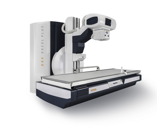
milla1cf Thu, 01/11/2024 - 08:22 January 11, 2024 — Carestream Health has launched a new and enhanced DRX-Excel Plus X-ray System that boosts the performance of the powerful, two-in-one solution to enable more productivity and efficiency, higher image quality, and an improved experience for users and patients.
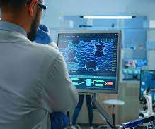
CHICAGO -- German researchers are testing ways to support nonradiologists in interpreting chest x-rays in emergency settings using an AI assistant. To test the AI algorithm, the LMU team focused on 563 cases, all ordered by the emergency unit department. Nonradiology residents 0.78
A study published in the Journal of Medical Radiation Sciences compared 270 dental X-rays, from people who were told to hold their tongues against the roof of their mouths throughout the procedure.
Teleradiology-India Introduction: “X-ray Visionaries” takes you on a compelling journey to unveil the expertise of radiographers and technologists, the unsung heroes of X-ray technology. The importance of minimizing radiation exposure while ensuring precision in diagnostic imaging.
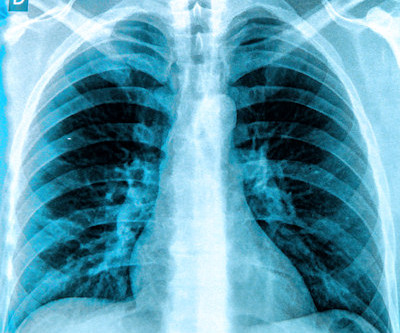
A fundamental goal of radiographers is to complete an imaging exam that provides sufficient information for an accurate clinical diagnosis–and at the lowest possible dose. To make it easier for readers, I’ve organized the available solutions into three exam types: general X-ray, chest imaging, and pediatric imaging.
In a new study, a radiograph device equipped with an AI-powered deep learning neural network has demonstrated improved image quality in pediatric X-rays.
Carestream Health has launched a new x-ray system called DXR-Excel Plus with features designed to help simplify workflow and enhance productivity. Carestream's new DXR-Excel Plus x-ray system. Image courtesy of Carestream Health.
The company will showcase the clinical analysis of Canon’s Intelligent Noise Reduction (Intelligent NR) that provides superior image quality while lowering radiation dosing in pediatric digital radiography at the Radiological Society of North America Annual Meeting 2023 (RSNA) , McCormick Place Convention Center, Chicago, IL Nov.
Detecting CSIs in a clinical setting often requires imaging such as X-rays and computed tomography (CT) scans, both of which expose children to radiation, which can cause other health issues over time.
Shamie Kumar describes how AI fits into a radiology clinical workflow and her perspective on how a clinical radiographer could use this to learn from and enhance their skills. If the AI findings are seen in PACS, how many radiographers actually log into PACS after taking a scan or X-ray? Can Radiographers Up-Skill?
milla1cf Fri, 02/23/2024 - 10:22 February 23, 2024 — The American Society of Radiologic Technologists (ASRT) launched its "Be Seen" campaign today to raise public awareness about the crucial role medical imaging and radiation therapy professionals play in patient diagnosis, intervention and treatment.
Lumbosacral spine X-rays, also called lumbar spine X-rays, are a radiographic imaging technique that uses low doses of electromagnetic radiation to view the internal anatomy of the lower spine, called the lumbosacral region.
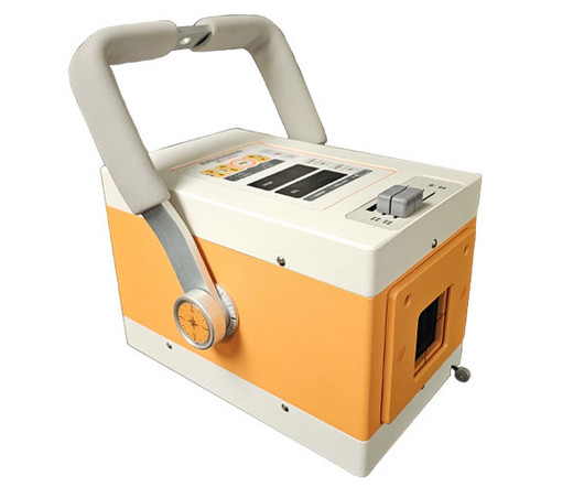
The Reveal Mobi Pro integrates KA Imaging’s Reveal 35C detector with SpectralDR technology into a complete mobile X-ray solution. The Reveal 35C detector mimics the workflow, dose, and techniques of state-of-the-art mobile DR X-ray. Our radiographic spectral images separate materials such as water (i.e.,
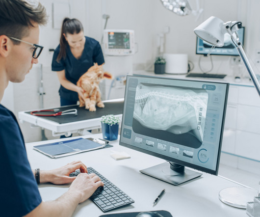
One such innovation that has transformed the way veterinarians approach imaging is the evolution of portable x-ray units. However, the advent of portable x-ray units has ushered in a new era of convenience and efficiency.
Common diagnostic tests for pulmonary disorders include chest x-rays and pulmonary function tests (PFTs). a) Raw example of a dynamic digital radiograph. (b) The digital technology limits radiation exposure to patients compared with standard chest x-rays, they wrote.
DDR is an emerging imaging technique that uses a pulsed x-ray source to acquire a series of radiographs at six to 15 frames per second. These images are then processed to visualize joints in motion.
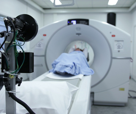
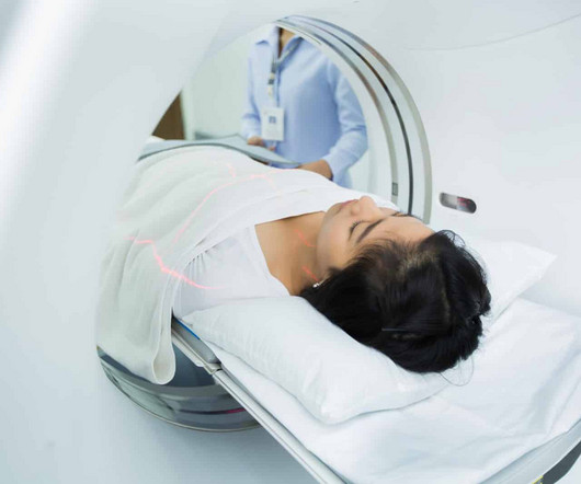
For X-rays, it usually takes less than 10 minutes. This article will talk about the different diagnostic imaging methods such as X-rays, CT scans, Ultrasound, and MRI. X-ray Also called a radiograph, an X-ray uses radiation to create images of the body.
Introduction: Dental X-ray technologists, also known as dental radiographers, are the unsung heroes behind the scenes of every successful dental diagnosis and treatment. In this blog, we’ll take you on a journey inside the jaw and explore a day in the life of a dental X-ray technologist.
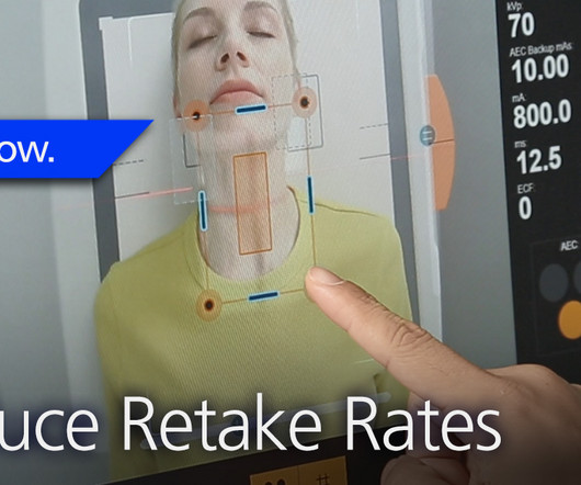
Repeating imaging exams increases the workload of your radiographers who are already stretched too thin; increases the exposure of the affected patients; and contributes to patients’ reduced confidence and satisfaction with your imaging department. The Audio Assist makes it easier for radiographers to hear the patients.
A patient’s specific needs and concerns are assessed, and a personalized radiographic plan is developed, taking into account factors like age, health conditions, and pregnancy. Minimizing Radiation Exposure: One of the paramount concerns in dental radiography is reducing radiation exposure.
Commercially Available Chest Radiograph AI Tools for Detecting Airspace Disease, Pneumothorax, and Pleural Effusion. Effects of low-dose ionizing radiation on genomic instability in interventional radiology workers. Generative Artificial Intelligence for Chest Radiograph Interpretation in the Emergency Department.
Spirometry values were below predicted values and a standard chest radiograph depicted an elevated right hemidiaphragm that was not present on a prior CT examination during his SARS-CoV-2 infection. With the dynamic functional imaging capability of DDR, the authors could visualize thoracic and pulmonary motion and track diaphragm movement.
In an open forum for radiographers at ECR 2024, Yi Xiang Tay, of Singapore University Hospital's radiography and diagnostic imaging department, shared his team's systematic review of the impact of imaging referral guidelines on patients and radiology services.
Veterinary technicians play a pivotal role in the radiographic process within veterinary practices. Poor positioning can lead to retakes, increased radiation exposure, and misdiagnoses. Double-Check Alignment : Confirm that the area of interest is correctly aligned with the x-ray beam.
At RSNA 2023, look for AI-driven systems that radiographers can use to help make patient positioning faster and more precise, and bring consistency to the process, all of which help improve image quality and reduce the need for retakes. Even the most skilled radiographers can fail to get positioning just right.
Although the word “radiology” sounds like it involves radiation, that is not always the case – for example, MRI (magnetic resonance imaging) and ultrasound do not use radiation in their medical imaging technologies. A radiographer is a medical professional who performs the scanning on patients.
Spirometry, specifically forced expiratory volume of air in 1 second (FEV1), is a prognostic marker in cystic fibrosis, and alongside chest x-ray findings is the primary method for assessing lung health in patients. Yet FEV1 may not always reflect the severity of the airway obstruction, they noted.
Kim Mason Kim Mason, an Audit and Research Radiographer for Mid Yorkshire Teaching Hospitals Trust, talks about their role as well as the value of radiographer engagement in research activities and how to get involved. In December, 1895, Wilhelm Röntgen would x-ray the hand of Anna Bertha Ludwig, his wife, using a photographic plate.
Geoffrey Rubin, MD, of the University of Arizona College of Medicine Tucson, offered session attendees an overview of key issues in the radiology workforce landscape, highlighting a 2024 consensus committee report from the American Society of Radiologic Technologists (ASRT) on the future of medical imaging and radiation therapy.
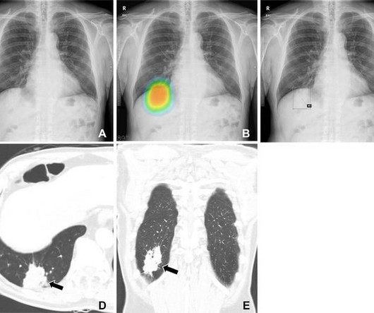
Researchers at Seoul National University looked at how these factors might influence the detection of malignant lung nodules during AI-assisted reading of chest X-rays. Of the 120 chest radiographs assessed, 60 were from lung cancer patients (32 males) and 60 were controls (36 males). Patients had a median age of 67 years.
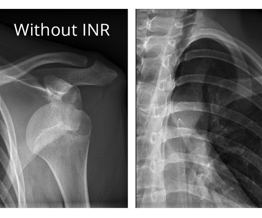
The technology of interventional X-ray systems has evolved. Today, the same should be said of AI ROI image-guided technology and the radiation reduction and protection it provides. Like pulsed fluoro before, AI ROI technology is proven to provide dramatic results in reducing radiation exposure – in reducing dose.
Not only are children more radiosensitive than adults (the cancer risk per unit dose of ionizing radiation is higher), but children also have a longer expected lifetime, which puts them at greater risk of cancer following radiation exposure.(1) The balance of dose and image quality is even more important in pediatric medical imaging.
Below is a description of each type of exam: Digital Mammography Mammography is the practice of radiographic imaging of the soft tissue of the breast. A mammogram is a low dose x-ray of the breast. There is no injection or radiation exposure associated with ultrasound.
Additionally, the organization reported it launched its ASRT “Be Seen” public awareness campaign in late February to raise awareness about the crucial role medical imaging and radiation therapy professionals play in patient diagnosis, intervention and treatment. ASRT 2024-2025 Board of Directors Daniel DeMaio, M.Ed., Marissa Mangrum, M.S.R.S.,
Although aimed at UK radiology trainees, it is also suitable for international residents taking similar examinations, postgraduate medical physics students and radiographers. The notes provide an excellent overview for anyone interested in the physics of radiology or just refreshing their knowledge.
It all started when Wilhelm Conrad Röntgen discovered X-rays in 1895. After working for weeks in his lab experimenting on the production of ‘strange rays’, which he referred to as ‘X’, he asked his wife Anna Bertha to lend ‘a hand’, the left one to be precise, which he used to produce the first X-ray image.
Closeup of X-ray photography of human brain Description: The field of radiology is in the midst of a remarkable revolution, driven by cutting-edge technologies and innovations. This description highlights the significant advancements in medical imaging that are transforming healthcare.
Frontal abdomen radiograph demonstrates foreign body consistent with capsule endoscopy device (pill cam) in descending colon. 5,6 ] Use of high-dose non-steroidal anti-inflammatory drugs, previous abdominal radiation therapy, and history of small bowel restrictions generally increase the risk of CR post-capsule endoscopy.
A normal lactate does not rule out the diagnosis Plain X-rays perform poorly in making or ruling out the diagnosis. A normal lactate does not rule out the diagnosis Plain X-rays perform poorly in making or ruling out the diagnosis. Abdominal X-ray 75% 66% 1.6 Absent bowel sounds Peritoneal signs (i.e.
Newborns' livers can be affected by a variety of congenital and acquired diseases, and imaging plays an important role in the workup and management of these, according to a study published November 7 in RadioGraphics. The team outlined the pros and cons of various modalities for neonatal liver imaging: X-ray. Ultrasound.
What if X-ray imaging, the most prevalent and accessible imaging modality in the world, could provide the information needed for diagnosis? This unique study will pair X-rays of consented patients with their high-resolution CTs (HRCTs) as well as weekly forced vital capacity (FVC) readings collected via an app for home spirometry.
We organize all of the trending information in your field so you don't have to. Join 5,000 users and stay up to date on the latest articles your peers are reading.
You know about us, now we want to get to know you!
Let's personalize your content
Let's get even more personalized
We recognize your account from another site in our network, please click 'Send Email' below to continue with verifying your account and setting a password.


Let's personalize your content