This site uses cookies to improve your experience. To help us insure we adhere to various privacy regulations, please select your country/region of residence. If you do not select a country, we will assume you are from the United States. Select your Cookie Settings or view our Privacy Policy and Terms of Use.
Cookie Settings
Cookies and similar technologies are used on this website for proper function of the website, for tracking performance analytics and for marketing purposes. We and some of our third-party providers may use cookie data for various purposes. Please review the cookie settings below and choose your preference.
Used for the proper function of the website
Used for monitoring website traffic and interactions
Cookie Settings
Cookies and similar technologies are used on this website for proper function of the website, for tracking performance analytics and for marketing purposes. We and some of our third-party providers may use cookie data for various purposes. Please review the cookie settings below and choose your preference.
Strictly Necessary: Used for the proper function of the website
Performance/Analytics: Used for monitoring website traffic and interactions
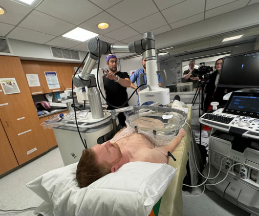
ORLANDO -- Sonographers on Earth can take a page or two from astronauts who use ultrasound in space, according to presentations given at the 2025 American Institute of Ultrasound in Medicine (AIUM) annual convention. Ultrasound has really helped space medicine quite a bit and will continue to expand.
Fluoroscopy-assisted ultrasound guidance for mini-percutaneous nephrolithotomy (mini-PCNL) procedures in children is a safer and more effective approach than fluoroscopy alone, researchers have found. But PCNL does impart radiation, and clinicians have sought to mitigate this exposure, especially to pediatric patients.
Contrast-enhanced ultrasound (CEUS) could be a suitable alternative to CT for early detection of complications in splenic trauma thats nonoperatively managed, according to a study presented on February 28 at ECR 2025. CEUS is a radiation-free method that can detect lesions within minutes using microbubble agents.
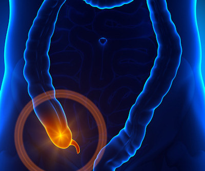
While the use of ultrasound for pediatric appendicitis imaging has increased, the modality -- as well as MRI -- remains underutilized in this area, according to research published August 22 in the Journal of Pediatric Surgery. Ultrasound alone was used in 53.3% Ultrasound alone was used in 53.3% NSQIP-P hospital 71.7%
A new, minimally invasive procedure that combines MRI and transurethral ultrasound is effective for treating prostate cancer, according to research presented March 20 at the Society of Interventional Radiology (SIR) meeting in Salt Lake City.
The use of more ultrasound translates to less CT use for diagnosing appendicitis in children, according to research published August 4 in the Journal of Surgical Research. These findings indicate that hospital experience in ultrasound and patient socioeconomic factors are correlated to overimaging,” Scaife and colleagues wrote.
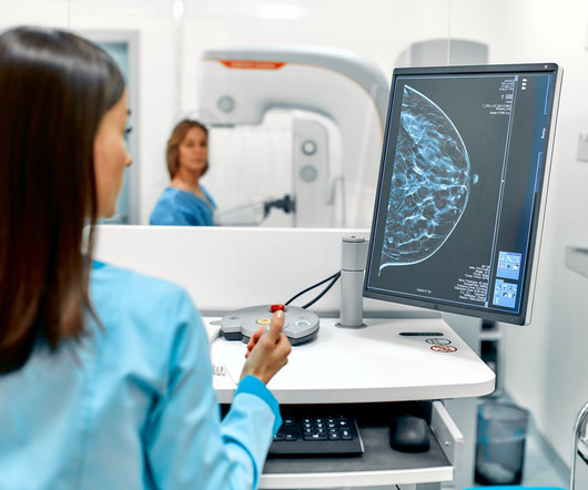
However, many patients have concerns about radiation exposure and the potential risks involved. The truth is mammograms are generally safe when used properly, and the amount of radiation you’re exposed to is minimal. Radiation exposure is controlled and minimized to ensure patient safety.
Ultrasound's utility will be on full display at this year's RSNA annual meeting, showing its merit in a wide variety of clinical applications. Studies will also evaluate ways to further improve the modality's capabilities by reassessing the physics side of ultrasound, such as attenuation and echogenicity, as well as the use of interferometry.
Studies to be presented in Chicago will explore ultrasound’s clinical applications in musculoskeletal, pediatric, abdominal, and women's imaging among other applications. Furthermore, ultrasound's use as a supplemental tool will be explored, including for breast cancer detection and follow-up imaging. And let’s not forget about AI.
Initial percutaneous nephrostomy (PCN) tube placement leads to more radiation exposure for pregnant women with suspected kidney stones, according to a study published October 27 in Urology. The Lyon team wanted to investigate the potential differences in radiation exposure per suspected stone episode between the options.
Gastrointestinal imagers should use high-quality MR enterography for complex inflammatory bowel disease (IBD) cases that can't be dealt with using intestinal ultrasound, even though both tests contribute to overall assessment, said an expert leading an advanced course on imaging Crohn's disease at ECR 2025. "MR
While services for breast and lung cancer screening were temporarily halted, imagers in x-ray, lung ultrasound, and PET/CT were busy examining patients who presented with COVID-19. He added that radiation treatment rooms are not completely sterile like operating rooms for surgeries. But that involves human contact.
Mammography: This technique is used to detect early signs of breast cancer and other abnormalities in breast tissue Low Dose CT for Lung Cancer Screening: This is a non-invasive imaging test that uses low levels of radiation to detect lung abnormalities early, improving the chances of successful treatment.
Dynamic contrast-enhanced ultrasound (CEUS) is a useful tool for interventions, according to a March 3 presentation given at ECR 2024. The team found that with ultrasound contrast, it could delineate the anatomy more clearly without linear signals from standard tissue. Pictured, he explains how it can better visualize endoleaks.
Previous studies on inappropriate imaging have focused on "the importance of minimizing patient radiation exposure, limiting patient discomfort, lost work time, use of resources efficiently, and reduction of overall healthcare expenditures," the group explained. to 23 kilotons for ultrasound. to 46 kilotons for x-ray, and 2.7
Cryoablation uses imaging guidance typically with ultrasound or CT to locate tumors. Previous research suggests that when the procedure is combined with hormonal therapy and radiation, patients can have nearly 100% of their tumors destroyed, Bryce noted. hormone therapy and radiation) therapies can have on this patient population.
Lymphosonography can detect sentinel lymph nodes (SLNs) in cervical and vulvar cancer patients scheduled for surgery, suggest findings presented March 30 at the 2025 American Institute of Ultrasound in Medicine (AIUM) annual conference.
Histotripsy uses focused ultrasound to deliver external beams intended to destroy cancerous tissue while leaving surrounding tissue intact. Histotripsy could become an option for patients who might otherwise be unlikely candidates for radiation or other ablation techniques, according to NYU Langone Health.
The organization noted that the CMS expects the overall impact of the proposed MPFS to be neutral on radiology, nuclear medicine, and radiation oncology. Centers for Medicare and Medicaid Services (CMS) proposed Medicare Physician Fee Schedule (MPFS) 2025 rule. However, interventional radiology will have a decrease of 2%.
In an effort to evaluate the appropriateness and outcomes of ultrasound, CT, and MR exams ordered for adult ED patients at a tertiary care urban academic center, the researchers retrospectively reviewed consecutive orders from January to March 2019. of ultrasounds, 29.1% of ultrasound orders, 61.4% of ultrasound orders, 61.4%
It uses electromagnetic radiation to examine bone structures to diagnose bone fractures, joint dislocations, and lung conditions. UltrasoundUltrasounds are more than a prenatal test offered to pregnant women; they can also be used to diagnose conditions affecting the abdomen and pelvis.
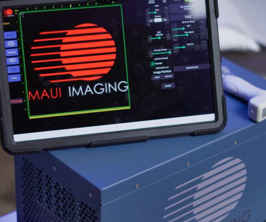
The trauma medicine project will incorporate MAUI's CET system, which the company describes as a cross between ultrasound and CT, which doesn't use ionizing radiation.
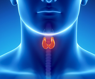
A deep learning-based framework incorporating ultrasound images and clinical data can help in thyroid nodule prediction, according to research published November 25 in the Journal of Radiation Research and Applied Sciences. The team developed predictive models using XGBoost, random forest, and support vector machines.
Lymphedema is a chronic disease of the lymphatic system caused by the accumulation of proteins in the interstitium, ultimately leading to inflammation, and can be caused by damage to the lymphatic system from surgery or radiation treatment, the authors explained. The disease is a particular concern among cancer patients, they noted.
Background: The increased utility and accessibility of point-of-care ultrasound (POCUS) has allowed clinicians the freedom to rethink their diagnostic approach for many common diseases, including peritonsillar abscess (PTA). Test characteristics of ultrasound for the diagnosis of peritonsillar abscess: A systematic review and meta-analysis.
At UCSF Benioff Children's Hospitals in Mission Bay and Oakland, radiologists are now offering a new, more comfortable, and radiation-free imaging method called pediatric Contrast-enhanced voiding urosonography (ceVUS).
Ultrasound offers another screening tool and can overcome some of mammography's limitations. Screening with breast MRI is "recommended for very high-risk patients, such as BRCA mutation carriers, as they begin screening at a young age and may be more susceptible to the negative effects of ionizing radiation," the authors wrote.
CEUS uses ultrasound contrast agents comprised of suspensions of tiny injectable “microbubbles” that do not contain dye, create no known risk of kidney damage or deposit of contrast media in the brain, and do not expose patients or hospital staff to ionizing radiation. For more information: www.icus-society.or
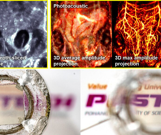
The 'ultrasound-photoacoustic dual-modal imaging system' combines molecular imaging contrast with ultrasound imaging, and it can visualize molecular and structural information inside the body in real time without any ionizing radiation.
She currently serves as chair of the department of medical imaging and radiation sciences in the College of Health Professions at TJU and directs the radiography and invasive cardiovascular technology programs. Her department currently serves 109 students who are studying in one of the university's nine concentration options.
Body imaging and radiation therapy technologists came in last, with annual base salaries of $73,000. Note: Salaries are for all U.S. regions and all healthcare organization types. The Mountain region -- eighth in radiologist salaries -- was at the top of this year's technologist salaries, at $133,000.
milla1cf Wed, 04/24/2024 - 19:18 April 24, 2024 — The International Contrast Ultrasound Society ( ICUS ) and Northwest Imaging Forums ( NWIF ) announced an educational partnership to help train sonographers to perform contrast-enhanced ultrasound (CEUS) examinations and administer intravenous ultrasound contrast agents.
Despite what its name implies, SRS isn’t actually a surgery but instead involves highly focused radiation that targets tumors while minimizing the effects on surrounding healthy tissue. The two-year meningeal disease rate was 5.8%, and the two-year symptomatic adverse radiation effect rate was 5%.
However, conventional CT requires hundreds of X-ray projections from multiple angles, exposing patients to significant radiation doses and relying on large, immobile systems. A New Paradigm in Imaging "In XACT, the generated sound waves by X-rays change the way X-ray imaging works, converting X-rays to ultrasound.
In these instances, contrast-enhanced ultrasound (CEUS) is rapidly becoming a viable alternative as an increasingly important tool for lesion characterization. Visit Philips EPIQ Elite for more information on Philips premium ultrasound. For more information: www.philips.com Reference: [1] Contract required.
Philips is introducing a new super contrast-enhanced ultrasound (CEUS) applicatio. Read more on AuntMinnie.com Related Reading: Philips issues radiation warning for fluoroscopy, angiography systems Philips begins U.S./European
Ultimately, compared with Tc-99m MIBI SPECT/CT, F-18 FCH-PET/CT offers a shorter scan time (about 90 minutes vs. at least 150 minutes), as well as significantly lower exposure to radiation, the researchers noted, and compared with neck ultrasound, F-18 FCH PET/CT identifies more hyperfunctioning glands.
Imaging techniques such as computed tomography, magnetic resonance imaging, positron emission tomography and ultrasound have become indispensable in the medical world. Each method not only opens unique insights into people's insides, but also allows physicians to draw conclusions about defects or functional processes in the human body.
By observing the consequences of the physical-chemical mechanisms at work within patients, this study shows that ultrasound elastography could be used in vivo as a quantitative indicator of the risk of breast implant rupture and help diagnose their replacement,” Ruffenach and colleagues wrote.
We organize all of the trending information in your field so you don't have to. Join 5,000 users and stay up to date on the latest articles your peers are reading.
You know about us, now we want to get to know you!
Let's personalize your content
Let's get even more personalized
We recognize your account from another site in our network, please click 'Send Email' below to continue with verifying your account and setting a password.


Let's personalize your content