This site uses cookies to improve your experience. To help us insure we adhere to various privacy regulations, please select your country/region of residence. If you do not select a country, we will assume you are from the United States. Select your Cookie Settings or view our Privacy Policy and Terms of Use.
Cookie Settings
Cookies and similar technologies are used on this website for proper function of the website, for tracking performance analytics and for marketing purposes. We and some of our third-party providers may use cookie data for various purposes. Please review the cookie settings below and choose your preference.
Used for the proper function of the website
Used for monitoring website traffic and interactions
Cookie Settings
Cookies and similar technologies are used on this website for proper function of the website, for tracking performance analytics and for marketing purposes. We and some of our third-party providers may use cookie data for various purposes. Please review the cookie settings below and choose your preference.
Strictly Necessary: Used for the proper function of the website
Performance/Analytics: Used for monitoring website traffic and interactions
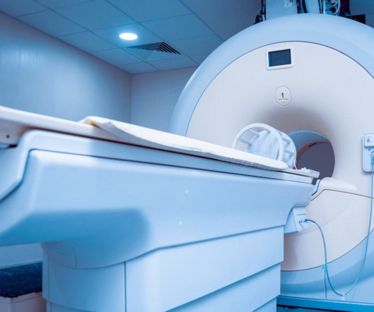
This year’s trip along the Road to RSNA for digital x-ray features a few familiar mileposts – AI for chest x-ray studies, for instance – but notably also significant research into how technology and new techniques can reduce radiation exposure in patients. Is adjacent x-ray imaging in the ER justified?
PT off the coast of Oceanside, CA, counting among its accomplishments the first use of an x-ray machine in space. Because of spectral technology for the first time you can do quantitative imaging with x-ray, Karim said. Stay tuned for part III, an interview with MinXRay , the portable x-ray machines developer.
A group in Japan has developed and tested an elastic x-ray shield made by embedding bismite particles in porous polyurethane, and suggests the new shielding material could provide more comfortable radiation protection. “It Then, it was air-dried to fix the metal particles in the porous polyurethane.
This year’s trip along the Road to RSNA for digital x-ray features mileposts mostly set by AI research. Models will be proposed for applications ranging from predicting bone density on chest x-rays to generating complete reports on anterior cruciate ligament (ACL) tears. Nonetheless, AI is poised to take top headlines.
X-rays are a common component of diagnostic testing and industrial monitoring, used for everything from monitoring your teeth to scanning your suitcase at the airport. But the high-energy rays also produce ionizing radiation, which can be dangerous after prolonged or excessive exposures.
Mercedes-Benz said it has performed the world's first crash test with a real car using x-ray imaging technology. The linear accelerator generates a continuous stream of x-ray pulses up to 1,000 images per second, which is about 1,000 times as many as with conventional medical x-ray procedures, the company noted.
The results suggest that there's a way to treat children with kidney stones with lower radiation doses, wrote a team led by Amr Salama, MD, of the Alexandria School of Medicine in Egypt. But PCNL does impart radiation, and clinicians have sought to mitigate this exposure, especially to pediatric patients. Hospital stay (days) 2.8
CHICAGO -- German researchers are testing ways to support nonradiologists in interpreting chest x-rays in emergency settings using an AI assistant. To test the AI algorithm, the LMU team focused on 563 cases, all ordered by the emergency unit department. Nonradiology residents 0.78
Canadian x-ray manufacturer KA Imaging and the charity Kenyan Kids Foundation Canada (KKFC) are collaborating to bring advanced medical imaging technology to Kenya. The project has an estimated cost of $100,000, which includes Reveal 35C, the x-ray source, software – including AI software for triage – and training.
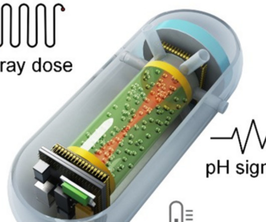
mm, the X-ray dosimeter measures radiation dose up to five times more accurately than standard methods. Coming in at a diameter of 5 mm and thickness of 0.2 mm,
X-rays are like a superpower for doctors—they can see things hidden from plain sight. Whether it’s a broken bone, a lung infection, or even a swallowed object, X-rays reveal the secrets of your body. At Professional Radiology in El Paso, we understand the importance of X-rays in guiding personalized care.
Mirion Dosimetry Services has announced commercial availability of a wearable radiation monitoring x-ray badge for medical imaging personnel and others. It also has a battery life of five years.
While services for breast and lung cancer screening were temporarily halted, imagers in x-ray, lung ultrasound, and PET/CT were busy examining patients who presented with COVID-19. He added that radiation treatment rooms are not completely sterile like operating rooms for surgeries. But that involves human contact.
S4-SSPH02-3 | Room S404 In this session on physics in radiography, a study suggests portable x-ray systems outfitted with digital autoexposure control (DAEC) systems enhance image quality and reduce radiation doses. Sunday, December 1 | 1:20 p.m.-1:30 1:30 p.m. |
The finding highlights the technique’s ability to reduce radiation doses in immunocompromised patients, noted lead author Ruxandra-Iulia Milos, MD, of the Medical University of Vienna in Austria, and colleagues. “To Currently, standard low-dose CT scans and chest x-rays are the preferred imaging methods for surveilling these patients.
However, conventional CT requires hundreds of X-ray projections from multiple angles, exposing patients to significant radiation doses and relying on large, immobile systems. A New Paradigm in Imaging "In XACT, the generated sound waves by X-rays change the way X-ray imaging works, converting X-rays to ultrasound.
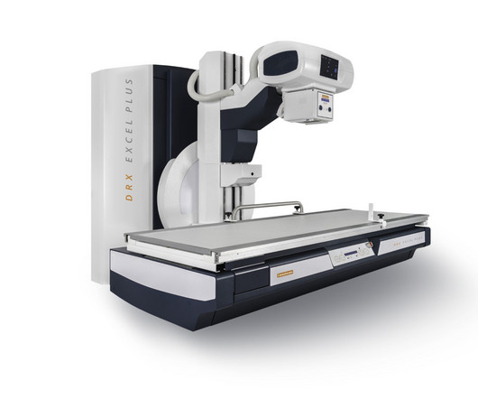
Carestream Health has launched a new x-ray system called DXR-Excel Plus with features designed to help simplify workflow and enhance productivity. Carestream's new DXR-Excel Plus x-ray system. Image courtesy of Carestream Health.
X-rays are standard procedures performed to diagnose and assess a variety of conditions or health concerns. What is the role of X-rays in medicine and how do they help improve people’s overall health? What is the role of X-rays in medicine and how do they help improve people’s overall health?
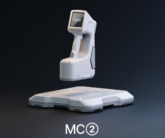
Food and Drug Administration (FDA) for its MC2 ultraportable x-ray system. MC2s small scatter area and low radiation output can reduce the space and infrastructure needs required by larger systems, the company said. It features a patented positioning system designed to assist in alignment for image capture.
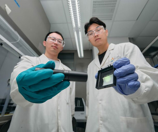
Not only could the materials reduce patient exposure to ionizing radiation, they also could reduce costs associated with traditional X-ray equipment, according to newly published research in Nature Communications.
Previous studies on inappropriate imaging have focused on "the importance of minimizing patient radiation exposure, limiting patient discomfort, lost work time, use of resources efficiently, and reduction of overall healthcare expenditures," the group explained. to 46 kilotons for x-ray, and 2.7 to 23 kilotons for ultrasound.
Canadian x-ray manufacturer KA Imaging and portable x-ray developer MinXray will assist with medical x-ray in space research via a project called SpaceXray.
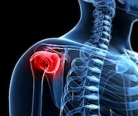
X-RaysX-Rays are one of the most commonly and widely used diagnostic imaging techniques. It uses electromagnetic radiation to examine bone structures to diagnose bone fractures, joint dislocations, and lung conditions. It is simple and quick to use, often taking less than 30 minutes to complete.
is powered by Fiber Optic RealShape (FORS) technology and allows doctors to navigate through blood vessels by using light rather than x-ray. The company added that LumiGuide's radiation-free technology could make way for better treatment of complex aortic procedures. Image courtesy of Philips.
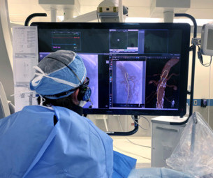
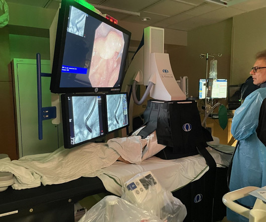
Carle Foundation Hospital installs innovative new technology that reduces the radiation risk to its endoscopy staff and patients. from Omega Medical Imaging is the most advanced interventional X-ray system available for endoscopy and the only fluoro system proven to reduce radiation exposure by up to 84%.
Researchers are developing novel scintillation materials for X-ray imaging applications that exhibit chemical stability, pose no toxicity risks, and can.
Canadian x-ray manufacturer KA Imaging plans to highlight clinical results regarding the use of its Reveal 35C detector in the intensive care unit (ICU) at the American Society of Emergency Radiology (ASER) meeting to be held in Washington, D.C.
milla1cf Thu, 09/21/2023 - 16:06 September 21, 2023 — Scientists in Moscow have successfully engineered a prototype detector for X-ray and PET/CT mаchines, using the perovskite-based photoconverters. These materials possess the unique ability to transmute radiation into an electric signal, yielding precise and illuminating images.
The American Society of Radiologic Technologists (ASRT) said it supports a new Pennsylvania bill that requires licensing of medical imaging professionals, radiation therapists, radiologist assistants, and trainees. Currently, there is no Pennsylvania state requirement for medical imaging professionals and radiation therapists to be licensed.
DDR is an emerging imaging technique that uses a pulsed x-ray source to acquire a series of radiographs at six to 15 frames per second. These images are then processed to visualize joints in motion.
. | M7-SSPH05-2 | Room N229 Findings will be presented in this Monday afternoon presentation on organ-specific ionizing radiation doses in neonatal patients who undergo interventional procedures for congenital heart disease (CHD). Gy-cm2, with organ-specific radiation doses highest for lung from frontal view (8.1
milla1cf Tue, 09/26/2023 - 15:32 September 26, 2023 — In a study of more than 2,000 chest X-rays , radiologists outperformed AI in accurately identifying the presence and absence of three common lung diseases, according to a study published in Radiology , a journal of the Radiological Society of North America ( RSNA ).
A point-of-care, web-based clinical decision support tool shows promise for not only reducing the incidence of inappropriate CT or MR imaging but also patients' radiation exposure and even carbon emissions caused by unnecessary exams, researchers have found. "[Our A reduction in effective radiation dose of 0.27 with 1 as reference).
Common diagnostic tests for pulmonary disorders include chest x-rays and pulmonary function tests (PFTs). The digital technology limits radiation exposure to patients compared with standard chest x-rays, they wrote. The study was published March 29 in Chest Pulmonary.
In a written statement, the organization leadership noted, “It can be argued that the discovery of the x-ray by German physicist Wilhelm Conrad Roentgen on November 8, 1895, was one of the most significant milestones in healthcare. Detectives reportedly used X-rays to determine if spouses were unfaithful.
A team of researchers at Boston Children’s Hospital has developed an age-specific dose catalog for estimating radiation exposure to children from diagnostic and interventional radiology fluoroscopy procedures.
Welcome to Maven Imaging's deep dive into C-arm radiation safety. As professionals in medical imaging, we understand how critical it is to keep your team and patients safe when operating digital X-ray equipment.
milla1cf Thu, 01/11/2024 - 08:22 January 11, 2024 — Carestream Health has launched a new and enhanced DRX-Excel Plus X-ray System that boosts the performance of the powerful, two-in-one solution to enable more productivity and efficiency, higher image quality, and an improved experience for users and patients.
A study published in the Journal of Medical Radiation Sciences compared 270 dental X-rays, from people who were told to hold their tongues against the roof of their mouths throughout the procedure.
"Because children are generally more prone to movement during scan acquisition than are adult patients, pediatric CT protocols tend to prioritize rapid image acquisition to reduce motion artifacts, in addition to radiation dose reduction," the group noted. Reduced radiation dose. Dual-source CT [i.e., Improved iodine signal.
SACRAMENTO, CA - A California radiologic technologist accused of delivering a major radiation overdose to a 23-month-old boy faced testimony this week on her role in the incident. There, a doctor ordered x-rays and CT scans to check for damage to the child's cervical spine. Is this safe?"
We organize all of the trending information in your field so you don't have to. Join 5,000 users and stay up to date on the latest articles your peers are reading.
You know about us, now we want to get to know you!
Let's personalize your content
Let's get even more personalized
We recognize your account from another site in our network, please click 'Send Email' below to continue with verifying your account and setting a password.


Let's personalize your content